
Fig. 2
- ID
- ZDB-IMAGE-080529-2
- Publication
- Sapede et al., 2005 - Role of SDF1 chemokine in the development of lateral line efferent and facial motor neurons
- All Figures
- Figures for Sapede et al., 2005
|
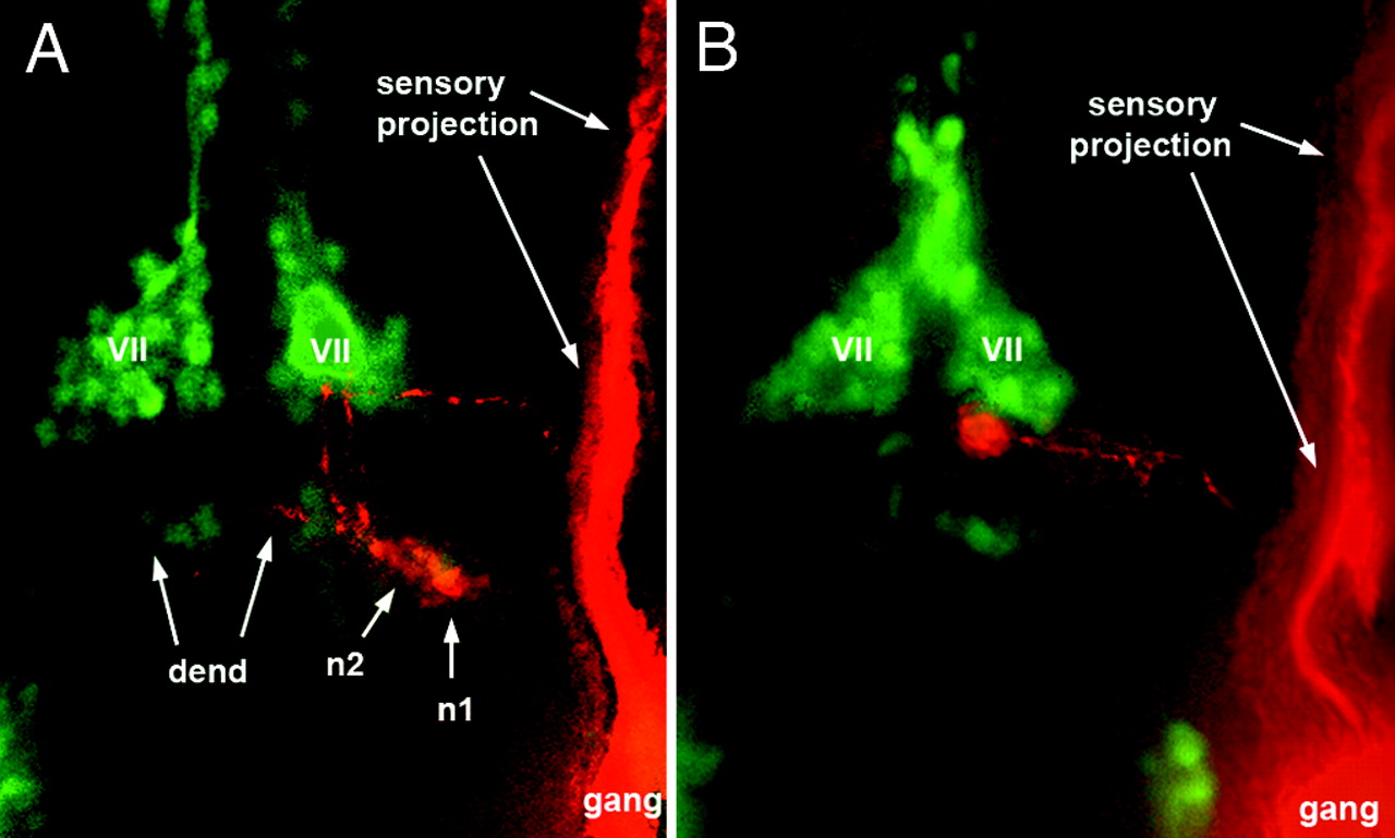
Fig. 2 Localization of caudal PLL efferent neurons relative to the facial motor nucleus as visualized in islet-GFP embryos. (A) Two efferent neurons, n1 and n2, occupy normal positions in a 3-day-old larva. GFP labeling is in green, DiI labeling is in red. (B) in a 48-haf embryo, the cell body is located at the posterior edge of the facial nucleus (VII) and has not yet reached its final, more posterior, position. The labeled efferent neurons appear red rather than yellow because the islet-GFP labeling was relatively faint and because the DiI labeling was enhanced to allow visualization of the axon (and partly of the dendrite, dend). gang, Ganglion.