Image


                        Figure Caption


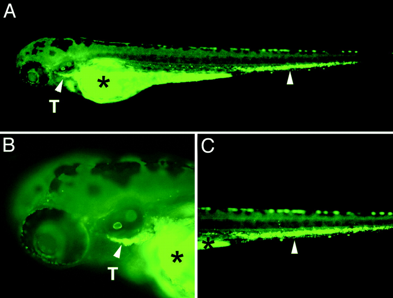
Fig. 4 GFP-labeled thymic T cells obtained from adult lck-GFP transgenic fish home to the thymus of transplanted embryos. (A) GFP fluorescent microscopic images of a 4-day-old transplanted embryo (anterior to the left and dorsal to the top) are shown. (B and C) Enlarged views of the head and tail region, respectively. Arrowheads denote GFP-labeled cells in the thymus (T) and in the tail region. Asterisks denote autofluorescence of the yolk sac.
                
                    
                        Acknowledgments


This image is the copyrighted work of the attributed author or publisher, and
ZFIN has permission only to display this image to its users.
Additional permissions should be obtained from the applicable author or publisher of the image.
      Full text @ Proc. Natl. Acad. Sci. USA


        