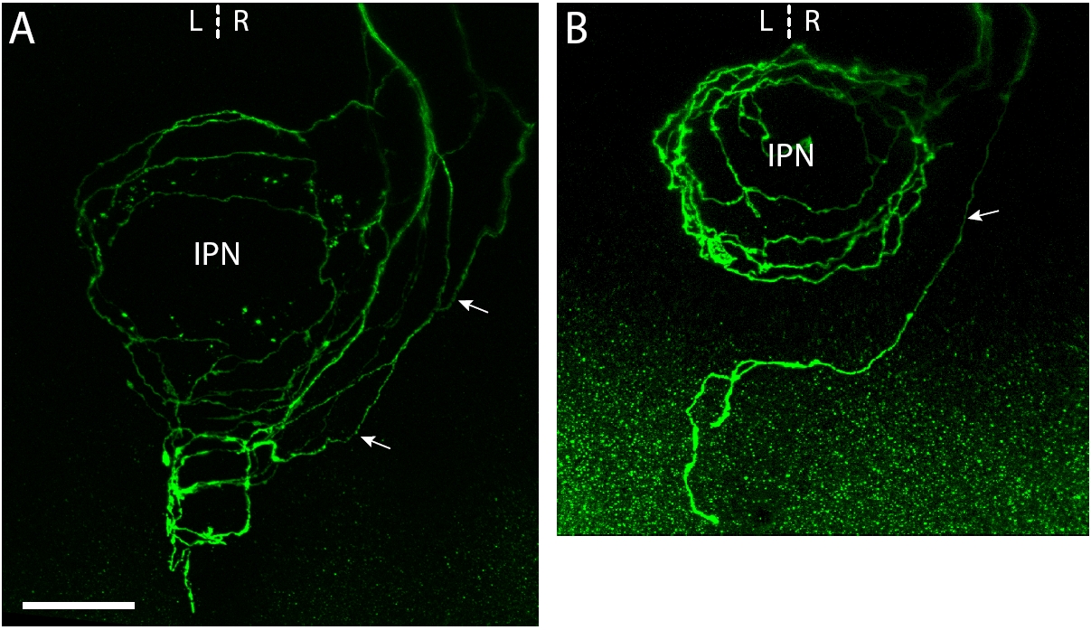
Fig. S1 A subset of habenular projection neurons extend axons that pass around the IPN and terminate in the anterior hindbrain. (a, b) Confocal z-projections of the ventral midbrain and anterior hindbrain in 5 dpf (a) and 8 dpf (b) larvae in which groups of habenular neurons have been labeled by focal electroporation. Some habenular neurons project axons that course ipsilaterally around the IPN (arrows) before converging medially to terminate on either side of the midline. These caudal terminations lie in the anterior hindbrain at the level of the serotoninergic raphé nucleus [16]. Scale bar: 25 μm.
Image
Figure Caption
Acknowledgments
This image is the copyrighted work of the attributed author or publisher, and
ZFIN has permission only to display this image to its users.
Additional permissions should be obtained from the applicable author or publisher of the image.
Full text @ Neural Dev.

