
Fig. 3
- ID
- ZDB-IMAGE-080522-9
- Publication
- Bernhardt et al., 2000 - Chondroitin sulfates affect the formation of the segmental motor nerves in zebrafish embryos
- All Figures
- Figures for Bernhardt et al., 2000
|
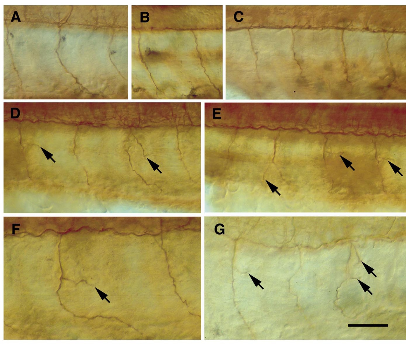
Fig. 3 Ventral motor nerves are revealed by immunostaining for acetylated α-tubulin in control (A–C) and in experimental embryos (D–G). Lateral views of whole-mount preparations, anterior is to the left, dorsal to the top. Arrows indicate abnormal side branches of the ventral motor nerve. (A) Ventral motor nerves (somites 7–9, left) extend without side branches along a midsegmental pathway in an uninjected embryo (26 hpf). (B) Anti-tubulin staining does not reveal side branches at least up to 36 hpf in uninjected embryos (shown here are ventral motor nerves 8 and 9, left). (C) Ventral motor nerves (7–10, right) are unbranched in an embryo (26 hpf) injected with vehicle solution. (D) Abnormal side branches have formed in two ventral motor nerves (8 and 10, left) in an embryo (26 hpf) injected with chondroitinase ABC (Sigma). (E) Same embryo as in (D), abnormal side branches have formed in three ventral motor nerves (8 –10, right). (F) Abnormal side branch in a ventral motor nerve (10, left) in an embryo (26 hpf) injected with chondroitinase ABC (Sigma). (G) Abnormal side branches in two ventral nerves (9 and 11, right) in an embryo (26 hpf) injected with chondroitinase ABC (Sigma). Magnification bar: 50 μm for (A–E), 30 μm for (F and G).
Reprinted from Developmental Biology, 221(1), Bernhardt, R.R. and Schachner, M., Chondroitin sulfates affect the formation of the segmental motor nerves in zebrafish embryos, 206-219, Copyright (2000) with permission from Elsevier. Full text @ Dev. Biol.