
Fig. 7
- ID
- ZDB-IMAGE-080516-37
- Genes
- Publication
- Qu et al., 2008 - ndrg4 is required for normal myocyte proliferation during early cardiac development in zebrafish
- All Figures
- Figures for Qu et al., 2008
|
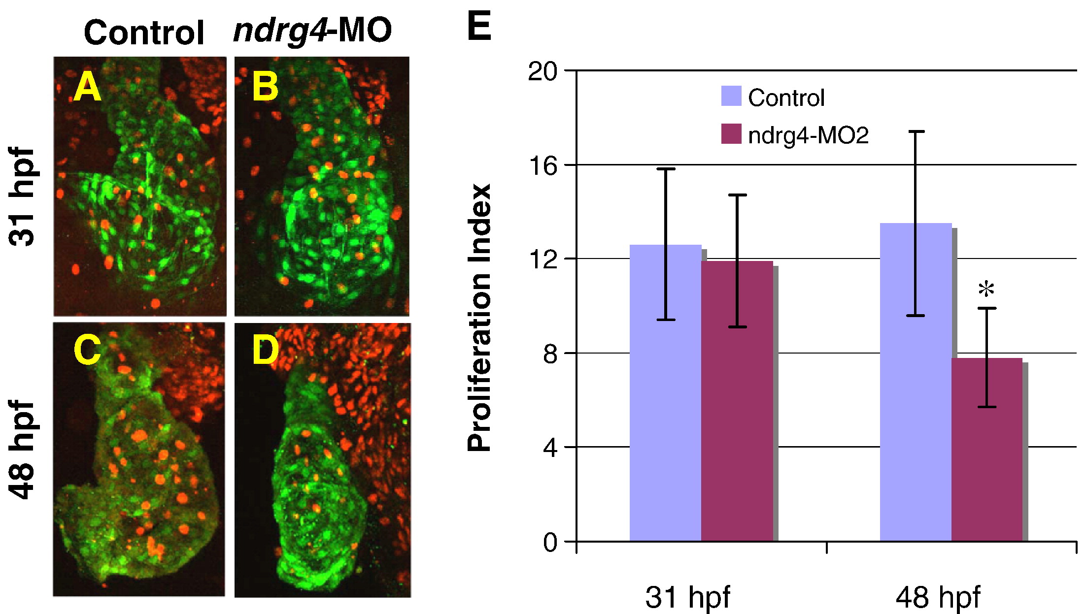
Fig. 7 Attenuation of Ndrg4 induces a reduction in cell proliferation of the cardiomyocytes. Although no significant difference in the number of BrdU positive cells between control (A) and ndrg4-MO2 treated (B) embryonic hearts at 31 hpf has been detected, ndrg4-MO2 injected embryos (D) display lower number of proliferating cells than control (C) in the heart at 48 hpf. All images represent reconstructions of confocal Z-stack sections imaged on whole hearts. Green, anti-GFP antibody; Red, anti-BrdU antibody. (E) Histogram showing proliferation index (the average of the total number of BrdU positive cells in per 100 cardiomyocytes) in the heart of 48 hpf embryos: control, 13.5 ± 3.9; ndrg4-MO2, 7.8 ± 2.1 (*P < 0.01).
Reprinted from Developmental Biology, 317(2), Qu, X., Jia, H., Garrity, D.M., Tompkins, K., Batts, L., Appel, B., Zhong, T.P., and Baldwin, H.S., ndrg4 is required for normal myocyte proliferation during early cardiac development in zebrafish, 486-496, Copyright (2008) with permission from Elsevier. Full text @ Dev. Biol.