
|
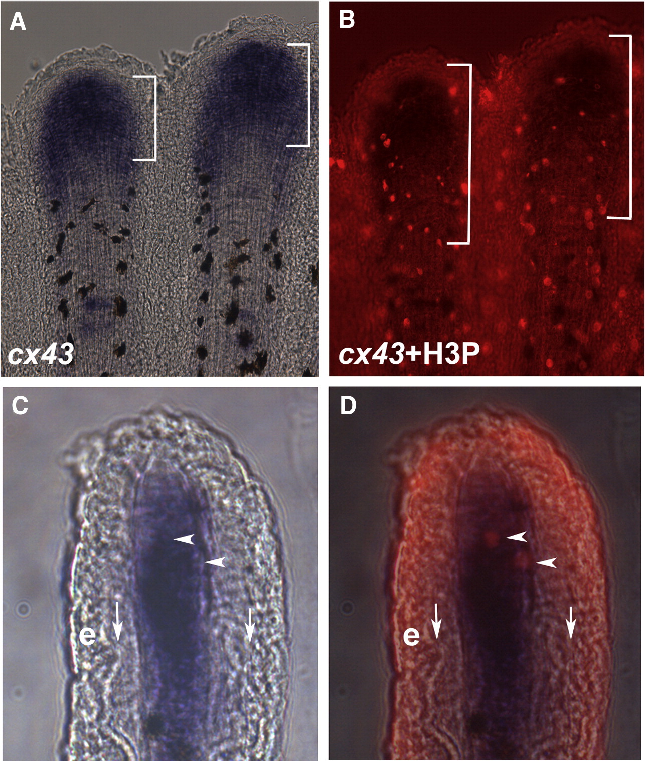
Fig. 2 cx43 is expressed in mitotic cells. Regenerating fins were labeled for both cx43 mRNA and H3P. (A) Whole mount in situ hybridization shows cx43 expression (brackets). The cx43 positive compartment measured from 70–140 μm from the distal end of the fin. (B) Double-staining for H3P and cx43 in situ hybridization. A distance of 250 μm from the distal end of the fin is shown in brackets. (C) Brightfield image of a single cryosection showing cx43 expression in the distal mesenchymal compartment. (D) Brightfield plus fluorescence reveals that the two H3P positive cells in this section are coincident with cx43 positive cells. Vertical arrows point to bone matrix; e, epithelium; arrowheads point to doubly-labeled cells.
Reprinted from Developmental Biology, 317(2), Hoptak-Solga, A.D., Nielsen, S., Jain, I., Thummel, R., Hyde, D.R., and Iovine, M.K., Connexin43 (GJA1) is required in the population of dividing cells during fin regeneration, 541-548, Copyright (2008) with permission from Elsevier. Full text @ Dev. Biol.