
|
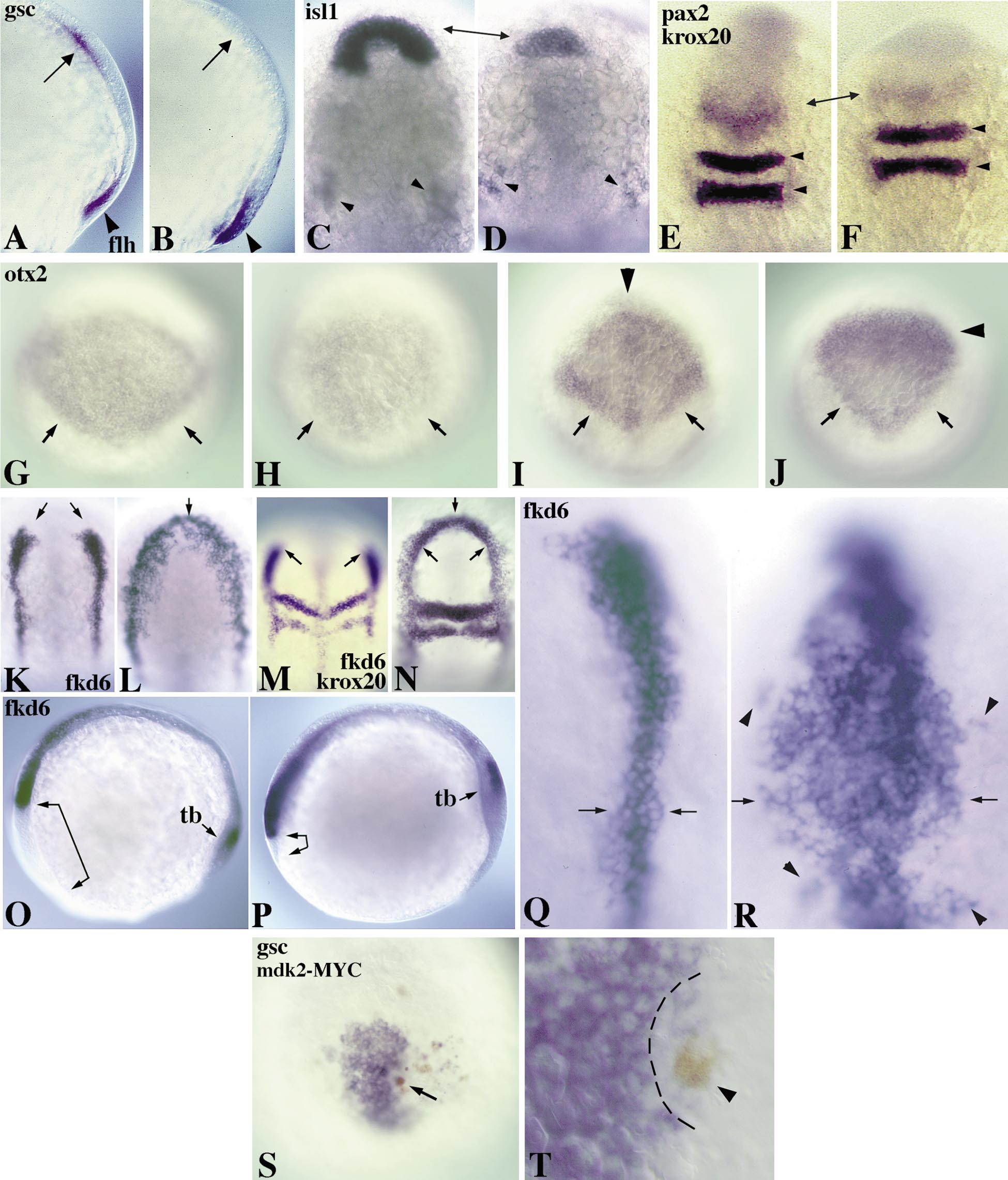
Fig. 3 Ectopic expression of mdk2 promotes posterior cell fates and blocks anterior development. (A, B) 80% epiboly embryos, lateral view. Expression of flh (also known as Znot; arrowhead) is unaffected in uninjected (A) and mdk2-injected embryos (B), but gsc (arrow) is repressed in B. (C–F) 12-h embryos, dorsal view, anterior to the top. (C) isl1 expression (arrows) in an uninjected embryo. (D) Repression of isl1 expression in the polster region of a mdk2 injected embryo (arrows). Trigeminal ganglion cells appear normal (arrowheads in C and D). (E) pax2 and krox20 expression in an uninjected embryo. (F) Reduction of pax2 expression in the MHB (arrows) in mdk2-injected embryo. Expression of krox20 in rhombomeres 3 and 5 (arrowheads) is unaffected. (G, H) 80% epiboly embryos, dorsal view, anterior to the top. (G) otx2 expression in the anterior neural plate of an uninjected embryo. Note pronounced expression at the caudal borders (arrows). (H) Reduction of otx2 expression in a mdk2-injected embryo. Border expression is lacking (arrows). (I–R) 12-h embryos, dorsal view (except O and P, lateral view). (I) Diamond-shaped expression of otx2 in the converging anterior neural plate with pronounced caudal borders (arrows) in an uninjected embryo. (J) Ectopic mdk2 expression results in a changed shape of otx2 expression with repression in the anterior domain (anterior to the arrowhead) and a reduction at the posterior edges (arrows). (K) fkd6 expression in premigratory neural crest cells of an uninjected embryo. (L) Expansion of fkd6-positive cells by ectopic expression of mdk2. This leads to a complete anterior fusion of the lateral fkd6 domains (arrows) and a substantial increase in the number of expressing cells. (M) Double labeling of fkd6 with krox20 in an uninjected embryo. (N) Anterior fusion of fkd6 domains (arrows). krox20 expression shows that anteroposterior register is not significantly changed. Lateral views (O, P) show mdk2-induced repression of anterior head structures (arrow-brackets) in mdk2-injected (P) versus uninjected (O) embryos. tb, tail bud. (Q, R) Higher magnification dorsal view of fkd6 expression in neural crest in uninjected (Q) and mdk2-injected (R) embryo. Arrows indicate the width of neural crest precursor populations, arrowheads show isolated population of crest cells. (S, T) 80% epiboly embryos, dorsal view. (S) Local repression of gsc expression in a DNA-injected embryo around single mdk2-MYC-expressing cell in the hypoblast (brown staining, arrow). (T) Higher magnification of a different embryo with repression of gsc in cells surrounding a mdk2-MYC-positive cell (brown, arrowhead). All embryos were injected with 200 pg RNA encoding mdk2 into 1 cell of the 2- to 4-cell stage embryo, except in S and T in which 20 pg DNA encoding MYC-tagged mdk2 was injected into 1 cell of the 8- to 16-cell stage embryo.
Reprinted from Developmental Biology, 229(1), Winkler, C. and Moon, R.T., Zebrafish mdk2, a novel secreted midkine, participates in posterior neurogenesis, 102-118, Copyright (2001) with permission from Elsevier. Full text @ Dev. Biol.