
Fig. 5
- ID
- ZDB-IMAGE-080514-45
- Publication
- Willot et al., 2002 - Cooperative action of ADMP- and BMP-mediated pathways in regulating cell fates in the zebrafish gastrula
- All Figures
- Figures for Willot et al., 2002
|
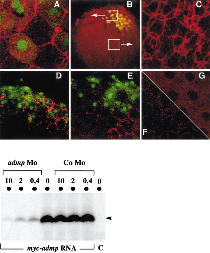
Fig. 5 Detection of the MYC-ADMP protein. (Top panel) myc-admp RNA and nls-gfp RNA (50 pg) were injected at the 16-cell stage into 1 marginal blatomere, and MYC-ADMP expression was revealed by immunocytochemistry at the sphere stage. NLS-GFP was used as a lineage tracer to identify the progeny of the injected cell. Embryos where observed with a confocal microscope. (A–C) myc-admp RNA (200 pg) (B)-injected embryo (10x objective); white boxes indicate a GFP-positive region and a GFP-negative region showed at a higher magnification in (A) and (C), respectively. (A, C, G) 63x objective. (A) Injected embryo, GFP-positive domain. (C) Injected embryo, GFP-negative domain. (G) Uninjected embryo. (D–F) 40x objective. (D–E) myc-admp (50 pg) and nls-gfp RNA (50 pg) (E) admp-morpholino (500μM) was injected at the one-cell stage prior to myc-admp and nls-gfp RNA injection at the 16-cell stage. (F) Uninjected embryo. (Bottom panel) myc-admp RNA (1μg) or no RNA (C) was translated in vitro in the absence (0) or in the presence of admp-morpholino (10, 2, or 0.4 μg in a reaction volume of 25 μl). The synthesis of MYC-ADMP protein (45 kDa, arrowhead) was inhibited by the admp-morpholino (admp-Mo) in a dose-dependent manner, and was not affected by the presence of a control morpholino (Co-Mo).
Reprinted from Developmental Biology, 241(1), Willot, V., Mathieu, J., Lu, Y., Schmid, B., Sidi, S., Yan, Y.-L., Postlethwait, J.H., Mullins, M., Rosa, F., and Peyriéras, N., Cooperative action of ADMP- and BMP-mediated pathways in regulating cell fates in the zebrafish gastrula, 59-78, Copyright (2002) with permission from Elsevier. Full text @ Dev. Biol.