
|
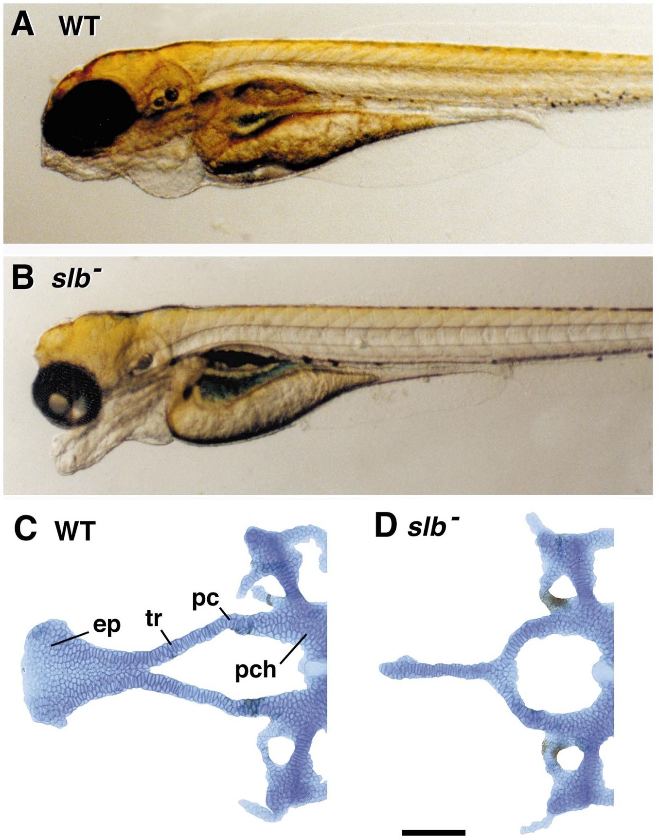
Fig. 3 The “bulldog-headed zebrafish,” comparing the wild-type (WT, A) and slb mutant (B) embryos at 2 days of development (unpublished photographs, courtesy of Dr. Corinne Houart). (C, D) Basicranial cartilages labeled with Alcian blue, dissected, and laid out as a flat mount, viewed from the dorsal aspect (unpublished, B. Ullmann and C.B.K.); pc, polar cartilage region; a distinctive region of joining between the parachordals and trabeculae. The polar cartilages form as separate elements in some organisms (e.g., see De Beer, 1937; Goodrich, 1930), but probably not in zebrafish. The abbreviations are as in Fig. 1.
Reprinted from Developmental Biology, 233(2), Kimmel, C.B., Miller, C.T., and Moens, C.B., Specification and morphogenesis of the zebrafish larval head skeleton, 239-257, Copyright (2001) with permission from Elsevier. Full text @ Dev. Biol.