
Fig. 9
- ID
- ZDB-IMAGE-080513-34
- Publication
- Isogai et al., 2001 - The vascular anatomy of the developing zebrafish: an atlas of embryonic and early larval development
- All Figures
- Figures for Isogai et al., 2001
|
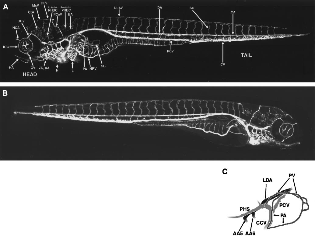
Fig. 9 Circulation in the developing zebrafish at approximately 4.5 days postfertilization (dpf). (A) Angiogram of a developing zebrafish at approximately 4.5 dpf, compiled from five separate reconstructions pasted together. Left-side lateral view, labeled. (B) Angiogram of a developing zebrafish at approximately 4.5 dpf, compiled from five separate reconstructions pasted together. Right-side lateral view, unlabeled. (C) Diagram illustrating the pectoral artery (PA) and pectoral vein (PV), and the source (from the DA) and drainage (into the ACV) of these vessels. This diagram was prepared from the angiographic stack used to generate panel (A). All panels are oriented with rostral to the left, and all lateral views are from the left side except for (B), which is a right-side view with rostral to the right. A glossary of the names corresponding to all labeled vessels is provided in Table 1.
Reprinted from Developmental Biology, 230(2), Isogai, S., Horiguchi, M., and Weinstein, B.M., The vascular anatomy of the developing zebrafish: an atlas of embryonic and early larval development, 278-301, Copyright (2001) with permission from Elsevier. Full text @ Dev. Biol.