
Fig. 1
- ID
- ZDB-IMAGE-080513-26
- Publication
- Isogai et al., 2001 - The vascular anatomy of the developing zebrafish: an atlas of embryonic and early larval development
- All Figures
- Figures for Isogai et al., 2001
|
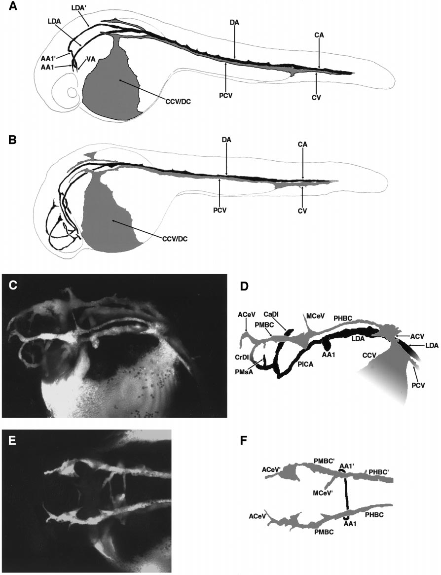
Fig. 1 Circulation in the developing zebrafish at 24–28 h postfertilization. (A) Diagram of active vessels in the zebrafish embryo just after the initiation of circulation, at approximately 24 h postfertilization (hpf). A single circulatory loop is present passing obligatorily through the trunk tail. (B) Diagram of active vessels in the zebrafish embryo a short time later, at approximately 26 hpf. A second cranial circulatory loop is now present. The stages in (A) and (B) are depicted using drawings because the weak circulation at these stages makes acquisition of attractive angiographic images difficult. These patterns are, however, representative of those seen in angiograms. (C) Angiogram of the head of a developing zebrafish at approximately 28 hpf. Dorsal–lateral view. (D) Diagram of vessels in (C). Only the vessels on the left side are shown, for simplicity. The major, primary arterial and venous vessels of the head are visible. (E) Angiogram of the head of a developing zebrafish at approximately 28 hpf. Dorsal view. Only the major primary venous vessels (PMBC, PHBC) are visible. The primary arterial vessels (LDA, PICA) are present but are directly underneath and obscured by the PMBC and PHBC in this view. (F) Diagram of vessels in (E). All panels are oriented with rostral to the left, and all lateral views are from the left side. A glossary of the names corresponding to all labeled vessels is provided in Table 1.
Reprinted from Developmental Biology, 230(2), Isogai, S., Horiguchi, M., and Weinstein, B.M., The vascular anatomy of the developing zebrafish: an atlas of embryonic and early larval development, 278-301, Copyright (2001) with permission from Elsevier. Full text @ Dev. Biol.