
|
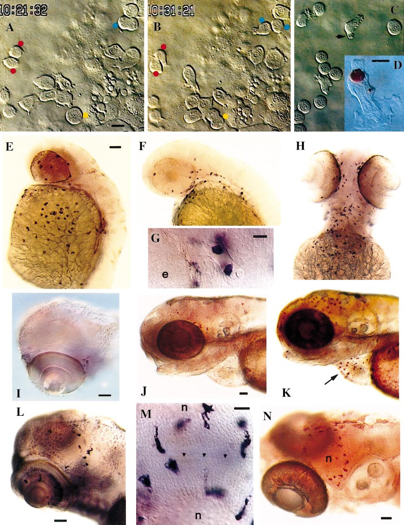
Fig. 9 Behavior of early macrophages in the panther mutant. All panther embryos, except K. (A–D) Live recordings of early macrophage behaviors in the yolk sac. (A, B) 26-somite stage (22 hpf). (A) Red dots mark sister cells from a mitosis that occured 15 min earlier; two other cells are presently in mitosis: the blue-dotted cell in anaphase B, the yellow-dotted one in pro-metaphase. (B) Ten minutes later, the blue-dotted cell cleaved in two daughter cells; the yellow-dotted cell is in anaphase. (C) 25 hpf; erythroblasts (rounded cells) have now joined the yolk sac and macrophages interact with them; one is phagocytosing an apoptotic residue (arrow). (D) 25 hpf, neutral red staining. (E–H) In situ hybridization for L-plastin. (E, F) Lateral views at 36 hpf (E; compare with wt in Fig. 2B) and 44 hpf (F). (G) 44 hpf, dorsal view, anterior to the left; two positive cells in a cephalic vessel. (H) 72 hpf, ventral view. (I, L) In situ hybridization for apoE at 72 hpf (I; compare with wild-type in Fig.7G) and 132 hpf (L); (J, K) 72 hpf; neutral red staining of live panther (J) and wild-type (K) embryos, showing macrophages associated with the optic tectum and regressing hatching gland (arrow). (M) 132 hpf; in situ hybridization for apoE, optic tectum, dorsal view, rostral to the left; (arrowheads) midline. (N) 7.5 days, neutral red staining (compare with wild-type at 99 hpf in Fig. 7F). (n) tectal neuropile. Bars: (A–C) 10 μm; (D) 5 μm; (E, F, H) 50 μm; (G) 10 μm; (I) 50 μm; (J, K) 50 μm; (L) 50 μm; (M) 20 μm; (N) 50 μm.
Reprinted from Developmental Biology, 238(2), Herbomel, P., Thisse, B., and Thisse, C., Zebrafish early macrophages colonize cephalic mesenchyme and developing brain, retina, and epidermis through a M-CSF receptor-dependent invasive process, 274-288, Copyright (2001) with permission from Elsevier. Full text @ Dev. Biol.