
Fig. 7
- ID
- ZDB-IMAGE-080513-15
- Publication
- Herbomel et al., 2001 - Zebrafish early macrophages colonize cephalic mesenchyme and developing brain, retina, and epidermis through a M-CSF receptor-dependent invasive process
- All Figures
- Figures for Herbomel et al., 2001
|
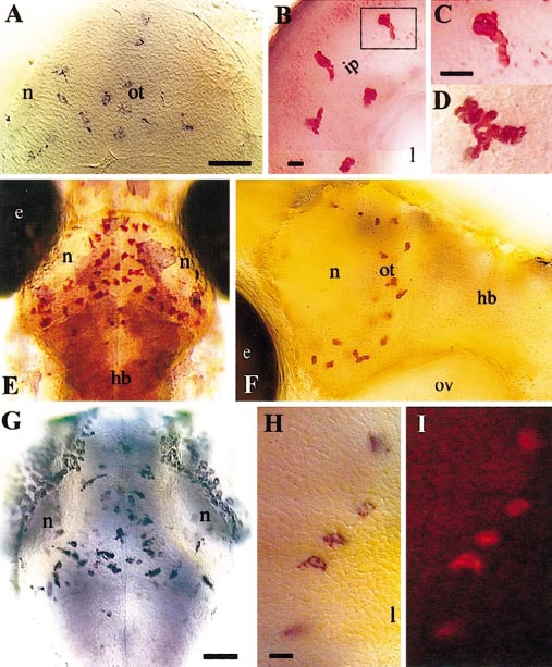
Fig. 7 Phenotypic transition of brain and retinal macrophages to early microglia; colonization of the optic tectum. (A, E, G) Dorsal view of the optic tectum, rostral upward. (A) 60 hpf, L-plastin in situ hybridization. (B–F) Neutral red-stained live embryos. (B) Retina, 65 hpf; the boxed early microglial cell is shown at higher magnification in (C). (D) Optic tectum, 82 hpf. (E, F) 99 hpf; (F) lateral view showing the extent of the left tectal neuropile, (G) In situ hybridization for apoE, 72 hpf. (H, I) Retina, 72 hpf; double in situ hybridization for L-plastin (blue) and apoE (red), (I) showing the red fluorescence of the apoE signal. (hb) hindbrain; (ot) optic tectum; (n) tectal neuropile; (e) eye; (ip) inner plexiform layer; (l) lens; (ov) otic vesicle. Bars: (A) 50 μm; (B) 10 μm; (C, D) 10 μm; (G) 50 μm; (H, I) 10 μm.
Reprinted from Developmental Biology, 238(2), Herbomel, P., Thisse, B., and Thisse, C., Zebrafish early macrophages colonize cephalic mesenchyme and developing brain, retina, and epidermis through a M-CSF receptor-dependent invasive process, 274-288, Copyright (2001) with permission from Elsevier. Full text @ Dev. Biol.