
Fig. 2
- ID
- ZDB-IMAGE-080513-11
- Publication
- Herbomel et al., 2001 - Zebrafish early macrophages colonize cephalic mesenchyme and developing brain, retina, and epidermis through a M-CSF receptor-dependent invasive process
- All Figures
- Figures for Herbomel et al., 2001
|
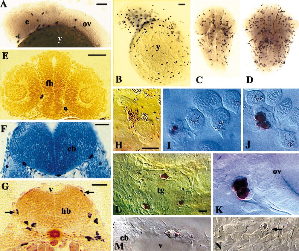
Fig. 2 Rapid spreading of early macrophages throughout the head. (A–G) In situ hybridization for L-plastin. (A) Lateral view of the head, 25 hpf. (B) Lateral view of the head and yolk sac, 35 hpf. (C, D) Dorsal views of the head at 35 hpf (C) and 48 hpf (D). (E–G) Cross sections; (E) 25 hpf; two macrophages in the interstices between eyes and forebrain (fb); (F) 35 hpf; three macrophages adhering to the ventral basal lamina of the cerebellum anlage (cb); (G) 35 hpf; macrophages caudal to the otic vesicle, migrating on the hindbrain walls (arrows) and inside the hindbrain ventrally. (H–N) Live embryos at 28–35 hpf viewed by DIC combined with neutral red staining (H–M), or by DIC alone (N); lateral views, dorsal up, anterior to the left. (H–J) Macrophages in the yolk sac blood circulation valley, showing the progression of the neutral red staining process (see text); cells with no stained lysosomes are erythroblasts. (K) Two probably sister macrophage cells adjacent to the otic vesicle. (L) Three macrophages associated with the trigeminal ganglion (tg). (M) Macrophage wandering on the roof of the fourth brain ventricle (v). (N) Phagocytic macrophage (arrow) among trigeminal ganglion neurons. (y) yolk sac; (e) eye; (ov) otic vesicle; (fb) forebrain; (hb) hindbrain; (cb) cerebellum anlage; (tg) trigeminal ganglion; (v) fourth brain ventricle. Bars: (A) 50 μm; (B–D) 50 μm; (E) 50 μm; (F, G) 25 μm; (H–K) 10 μm; (L–N) 10 μm.
Reprinted from Developmental Biology, 238(2), Herbomel, P., Thisse, B., and Thisse, C., Zebrafish early macrophages colonize cephalic mesenchyme and developing brain, retina, and epidermis through a M-CSF receptor-dependent invasive process, 274-288, Copyright (2001) with permission from Elsevier. Full text @ Dev. Biol.