
Fig. S3
- ID
- ZDB-IMAGE-080505-38
- Publication
- Bertrand et al., 2008 - CD41+ cmyb+ precursors colonize the zebrafish pronephros by a novel migration route to initiate adult hematopoiesis
- All Figures
- Figures for Bertrand et al., 2008
|
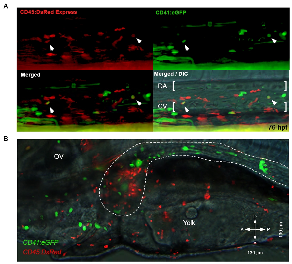
Fig. S3 A CD45:DsRed transgene marks a subset of AGM HSCs and differentiated leukocytes. (A) Deconvolved image stacks through the 76 hpf AGM shows a subset of CD45:DsRed+ cells (upper left panel) also express the CD41:eGFP transgene (upper left panel). Lower panels show merged channels, with Nomarski overlay on the right. Arrowheads mark double positive cells, brackets mark the boundaries of the dorsal aorta (DA) and cardinal vein (CV). (B) Double transgenic animals show GFP+ cells along the pronephric ducts and DsRed+ cells within the anterior pronephros. All animals oriented dorsal side upwards, anterior towards the left. OV, otic vesicle.