
IMAGE
Fig. 3
- ID
- ZDB-IMAGE-080505-31
- Publication
- Bertrand et al., 2008 - CD41+ cmyb+ precursors colonize the zebrafish pronephros by a novel migration route to initiate adult hematopoiesis
- All Figures
- Figures for Bertrand et al., 2008
Image
|
Figure Caption
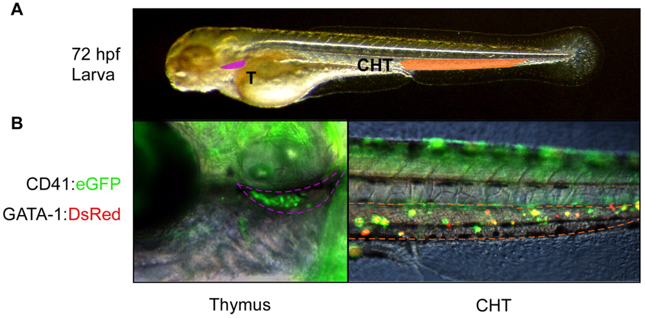
Fig. 3 Transplanted CD41:eGFP+ cells colonize the thymus and caudal hematopoietic tissues. (A) Photograph indicating regions shown at higher magnification in B (Nomarski/fluorescence merge). Purple region denotes left thymic lobe and orange region the CHT. (B) One day after transplantation, recipient animals showed robust colonization of thymi (left panel) and the CHT (right panel). Transplanted CD41+ cells also carried a gata1:DsRed transgene to visualize erythroid differentiation.
Acknowledgments
This image is the copyrighted work of the attributed author or publisher, and
ZFIN has permission only to display this image to its users.
Additional permissions should be obtained from the applicable author or publisher of the image.
Full text @ Development