
Fig. 2
- ID
- ZDB-IMAGE-080429-11
- Genes
- Publication
- Park et al., 2002 - olig2 is required for zebrafish primary motor neuron and oligodendrocyte development
- All Figures
- Figures for Park et al., 2002
|
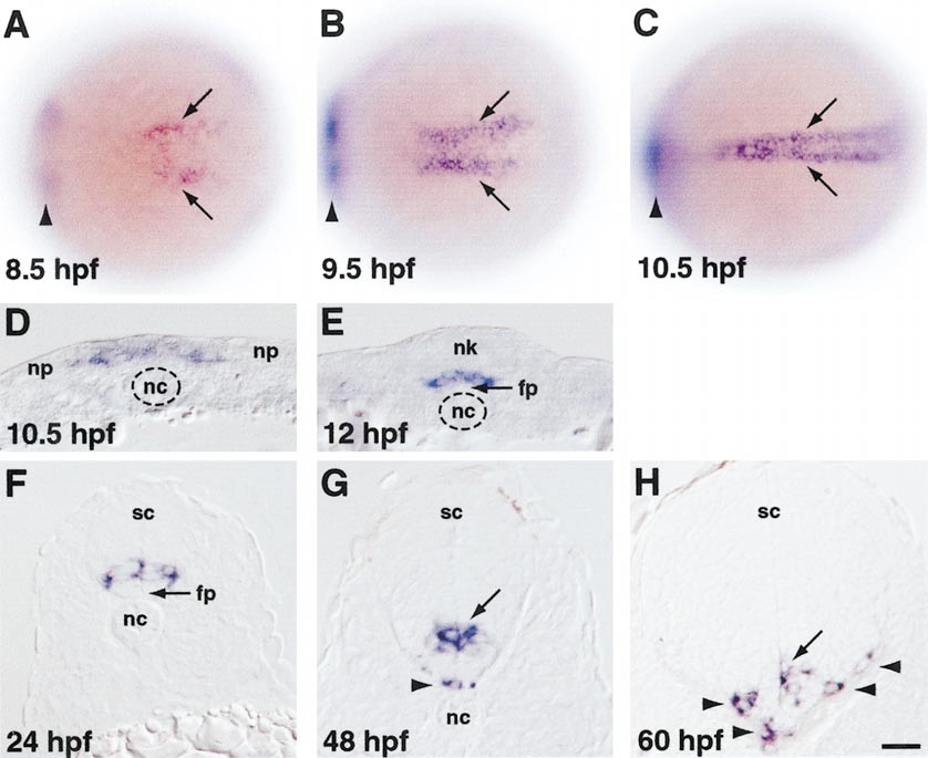
Fig. 2 olig2 expression revealed by in situ RNA hybridization. (A–C) Dorsal views, anterior to the left, of whole embryos. Arrowheads indicate expression in prospective ventral diencephelon, which is out of the focal plane. (A) Prospective trunk spinal cord cells begin to express olig2 at midgastrula stage (arrows). (B) By late gastrula stage, olig2-expressing cells form two longitudinal stripes bordering the embryonic midline. (C) At neural plate stage, olig2-expressing cells form a single longitudinal domain at the midline. (D–H) Transverse sections, dorsal to the top, through trunk region. (D) Medial neural plate (np) cells, overlying notochord (nc), uniformly express olig2. (E) As neurulation proceeds, olig2-expressing cells occupy ventral neural keel (nk), overlying floor plate (fp). (F) After neurulation, cells within the ventral portion of the spinal cord (sc) express olig2. (G,H) Some olig2-expressing cells occupy basolateral positions, associated with the white matter (arrowheads). Scale bar equals 80 μm for (A–C) and 20 μm for (D–H).
Reprinted from Developmental Biology, 248(2), Park, H.-C., Mehta, A., Richardson, J.S., and Appel, B., olig2 is required for zebrafish primary motor neuron and oligodendrocyte development, 356-368, Copyright (2002) with permission from Elsevier. Full text @ Dev. Biol.