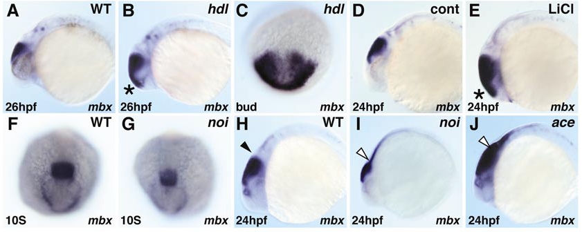
Fig. 3 mbx expression in mutant embryos. Whole-mount in situ hybridization of mbx. Black and white arrowheads indicate the MHB and putative MHB region, respectively. In headless (hdl) mutants, mbx expression is expanded rostrally at bud and 26-hpf stages (B, C). Similarly, anterior mbx expansion at 24 hpf is observed in embryos treated with 0.3 M LiCl just after shield stage (E). In the no isthmustu29a (noi) mutant, mbx expression is slightly diminished at the 10-somite stage (G), and reduced in intensity but expanded caudally at 24 hpf (I). In the acerebellarti282a (ace) mutant, mbx expression in the tectum is retained and again expanded caudally (J).
Reprinted from Developmental Biology, 248(1), Kawahara, A., Chien, C.-B., and Dawid, I.B., The homeobox gene mbx is involved in eye and tectum development, 107-117, Copyright (2002) with permission from Elsevier. Full text @ Dev. Biol.

