
Image
|
Figure Caption
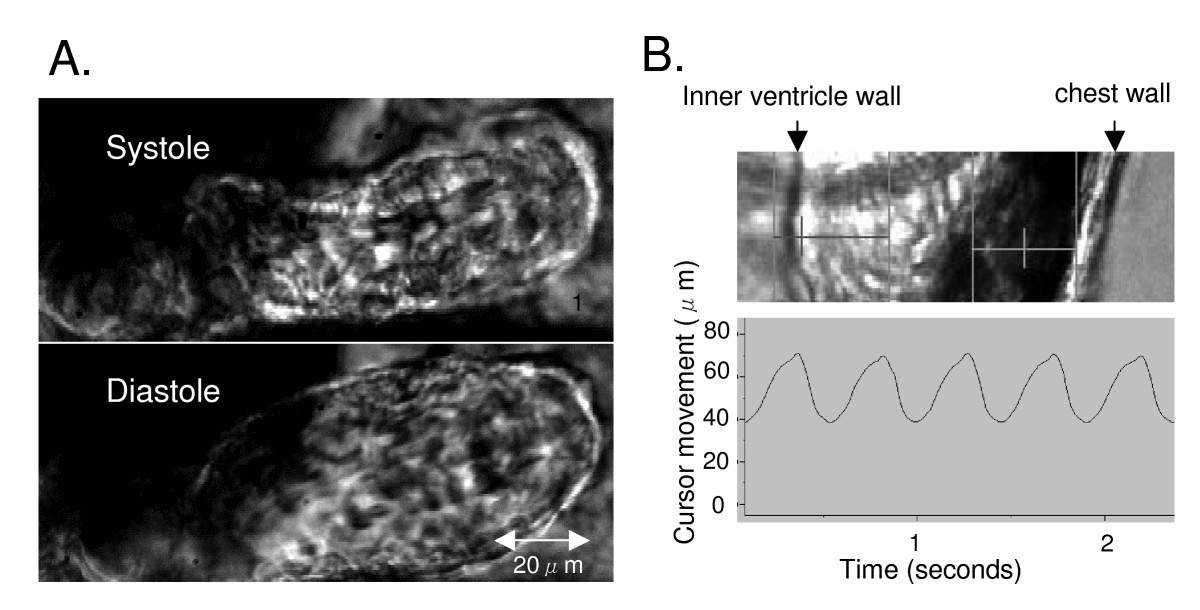
Fig. 3 2-day zebrafish embryo ventricle in systole and diastole (panel A). Inner ventricle wall is tracked by a cursor (panel B, left vertical line) and the movement displayed a continuous trace (panel B, contraction is upwards).
Acknowledgments
This image is the copyrighted work of the attributed author or publisher, and
ZFIN has permission only to display this image to its users.
Additional permissions should be obtained from the applicable author or publisher of the image.
Full text @ BMC Biotechnol.