
Fig. 7
- ID
- ZDB-IMAGE-080402-28
- Publication
- De Mazière et al., 2008 - Egfl7 knockdown causes defects in the extension and junctional arrangements of endothelial cells during zebrafish vasculogenesis
- All Figures
- Figures for De Mazière et al., 2008
|
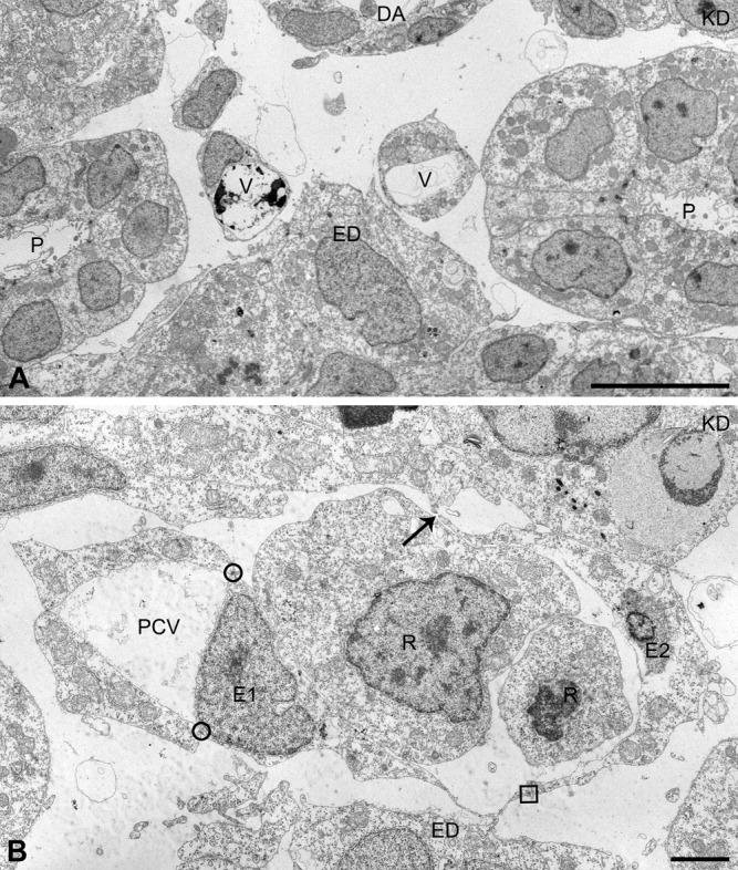
Fig. 7 Aberrant endothelial cell (EC) arrangements, different from vessel lumen obliteration, in the axial vascular area of the trunk of Egfl7 KD embryos at 24 hours postfertilization (hpf). In addition to reduced vessel lumens caused by cross-luminal EC-EC junctions, other types of structural defects were found in Egfl7 knockdown (KD) embryos. A: Example of a posterior cardinal vein (PCV) that splits into two small vessels, each associated with the endoderm. B: Example of a PCV with a small lumen and attached loose-ending (arrow) EC sheet. The EC denoted as E1 is arranged in the PCV endothelium by means of two normally positioned tight junctions (circles), and attached to the loose EC sheet (E2) by means of an aberrantly positioned tight junction (square) within the basal plasma membrane of E1. ED, endoderm; P, pronephric duct; R, red blood cell precursor; V, vein. Scale bars = 10 μm in A, 2 μm in B.