
Fig. 5
- ID
- ZDB-IMAGE-080326-42
- Publication
- Berghmans et al., 2005 - tp53 mutant zebrafish develop malignant peripheral nerve sheath tumors
- All Figures
- Figures for Berghmans et al., 2005
|
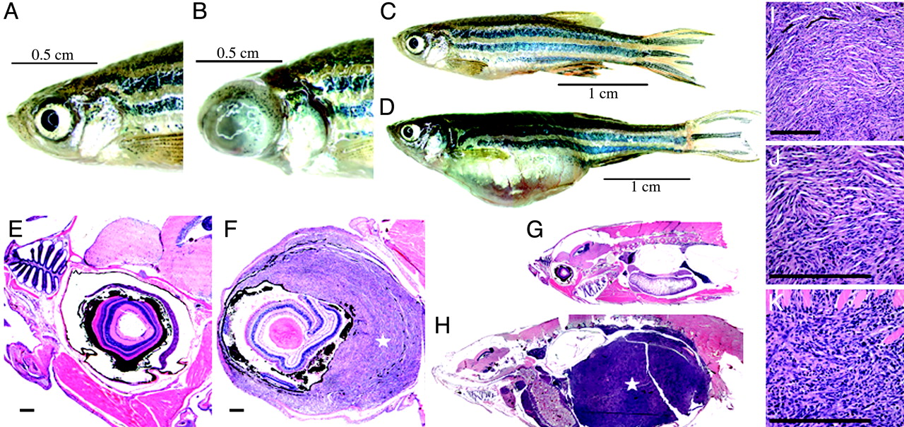
Fig. 5 Tumorigenesis features of tp53M214K mutant zebrafish. (A-D) When compared with wild-type zebrafish (A and C), zMPNST development in tp53 mutant zebrafish is identifiable upon external observation because of ocular (B) or abdominal tumor localizations (D). (E-H) When compared with wild-type zebrafish (E and G), histopathology staining with hematoxylin/eosin reveals zMPNST in the eye (F) and abdominal cavity (H), as indicated by the stars (x4). (I-K) Histopathological features of tumors (I) composed predominantly of spindle cells (J) and to a varying degree of epitheloid cells (K) are consistent with the diagnosis of zMPNST. [Bar, 200 μm (E, F, I-K).]