Fig. 4
Stra6 Deficiency Causes Multisystemic Malformations in Zebrafish
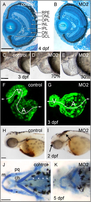
(A and B) Cross-sections through the eye of 4 dpf control (A) and MO2 morphant (B) larvae. The stra6 morphant eye is smaller but shows regular stratification of distinct retinal layers. L, lens; RPE, retinal pigment epithelium; ONL, outer nuclear layer; OPL, outer plexiform layer; INL, inner nuclear layer; IPL, inner plexiform layer; ON, optic nerve; GCL, ganglion cell layer.
(C–E) Close-up views of the pericardial region of control (C) and MO2-treated morphants (D and E). Characteristics of the heart shown in (D) represent 70% of the morphants; characteristics of the heart shown in (E) represent 30% of the morphants (n = 100). Note the absence of red blood cells in the heart due to a disrupted circulation in (E).
(F and G) Three-dimensional view of the heart of 3 dpf control (F) and MO2-treated (G) TG(fli1:EGFP)Y1 embryos. Blood flow is indicated by dashed lines. The ventricle (V) is on the left, and the atrium (A) is on the right.
(H and I) Control (H) and 2 dpf MO2-treated (I) embryos with the heart phenotype shown in (E) stained for hemoglobin. In controls, staining for erythrocytes was primarily detectable in the heart and afferent vessels over the yolk. In morphants, hemoglobin was greatly reduced in the heart and afferent vessels, but red blood cell extravasations were visible in the head (see arrows).
(J and K) Alcian blue staining of cartilage of the craniofacial skeleton of 5 dpf control (J) and MO2-treated morphant (K) larvae. The Meckel′s cartilage (m), palatoquadrates (pq), and ceratohyales (ch) were deformed and staining of ceratobranchials (asterisks) was highly reduced in Stra6 deficiency.
Animals were raised in PTU-free water. Anterior is to the left in all pictures. Scale bars = 100 μm in (A)–(E) and (H)–(K).
Reprinted from Cell Metabolism, 7(3), Isken, A., Golczak, M., Oberhauser, V., Hunzelmann, S., Driever, W., Imanishi, Y., Palczewski, K., and von Lintig, J., RBP4 Disrupts Vitamin A Uptake Homeostasis in a STRA6-Deficient Animal Model for Matthew-Wood Syndrome, 258-268, Copyright (2008) with permission from Elsevier. Full text @ Cell Metab.

