
Fig. 4
- ID
- ZDB-IMAGE-080326-18
- Antibodies
- Publication
- Smith et al., 2008 - Rotation and asymmetric development of the zebrafish heart requires directed migration of cardiac progenitor cells
- All Figures
- Figures for Smith et al., 2008
|
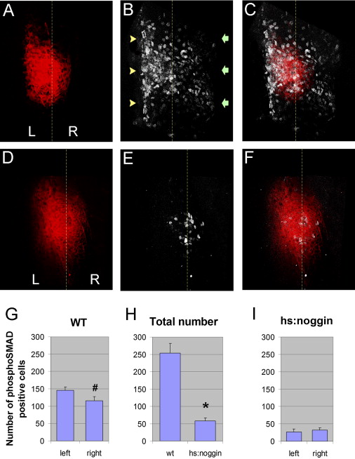
Fig. 4 Asymmetric Activation of Smad 1,5,8 Protein in the LPM. Confocal images of the LPM at the cardiac region in 23-somite stage embryos stained for tropomyosin (red, A and D), phospho-Smad 1,5,8 (white, B and E), and the overlay (C and F). Staining in WT embryos (A–C) shows asymmetric activation of Smad 1,5,8 on the left side (yellow arrowheads) compared with right (green arrows). Cell counting showed this difference to be statistically significant ([G]; mean ± SEM, # p < 0.05). Activation of Smad 1,5,8 was significantly lower in tg(hsp70:noggin3) embryos (D–F) compared with WT embryos ([H]; mean ± SEM, * p < 0.001), and the difference between left and right activation of Smad 1,5,8 was not significant ([I]; mean ± SEM). In all images anterior is to the top, left is to the left side, and the midline is demarcated by the dashed yellow line.
Reprinted from Developmental Cell, 14(2), Smith, K.A., Chocron, S., von der Hardt, S., de Pater, E., Soufan, A., Bussmann, J., Schulte-Merker, S., Hammerschmidt, M., and Bakkers, J., Rotation and asymmetric development of the zebrafish heart requires directed migration of cardiac progenitor cells, 287-297, Copyright (2008) with permission from Elsevier. Full text @ Dev. Cell