
Fig. 1
- ID
- ZDB-IMAGE-080326-15
- Genes
- Publication
- Smith et al., 2008 - Rotation and asymmetric development of the zebrafish heart requires directed migration of cardiac progenitor cells
- All Figures
- Figures for Smith et al., 2008
|
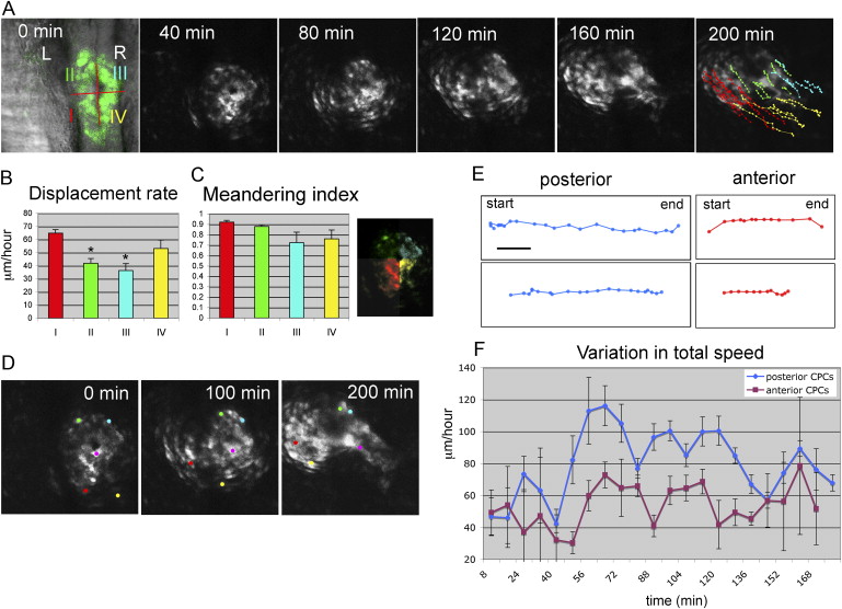
Fig. 1 Rotation of the Cardiac Cone during Formation of the Linear Heart Tube (A) Selected images of a confocal time-lapse recording of a tg(cmlc2:gfp) embryo starting at the 23-somite stage. Individual GFP-positive cells were tracked and color-coded according to their location within the cardiac field at the 23-somite stage. Dorsal view with anterior to the top. (B and C) Displacement rates and meandering index (displacement/track length) of CPCs located at different positions within the cardiac field (color corresponds to position indicated in the confocal image) calculated from time-lapse recording shown in (A) (n = 30). Error bars indicate standard errors. Statistic significance (by paired t test) calculated for CPCs derived from sector I (red) compared with CPCs derived from sectors II (green) and III (blue) (* p < 0.005). (D) Selected images of confocal timelapse recording with individual cells labeled during formation of the heart tube. Individual cells are marked (yellow, red, green, and blue for laterally located atrial cells and pink for medially located ventricular cells) and tracked over a 200 min period, revealing the evident rotation of the cardiac cone. (E) Representative tracks of individual cells originating from the posterior (blue) or anterior (purple) region of the cardiac cone with 8 min intervals in between time points shown. Scale bar represents 25 μm. (F) Graphical representation of CPC speeds during the process of heart tube formation. Cells were grouped according to their original position in the cardiac field (posterior, blue; anterior, purple).
Reprinted from Developmental Cell, 14(2), Smith, K.A., Chocron, S., von der Hardt, S., de Pater, E., Soufan, A., Bussmann, J., Schulte-Merker, S., Hammerschmidt, M., and Bakkers, J., Rotation and asymmetric development of the zebrafish heart requires directed migration of cardiac progenitor cells, 287-297, Copyright (2008) with permission from Elsevier. Full text @ Dev. Cell