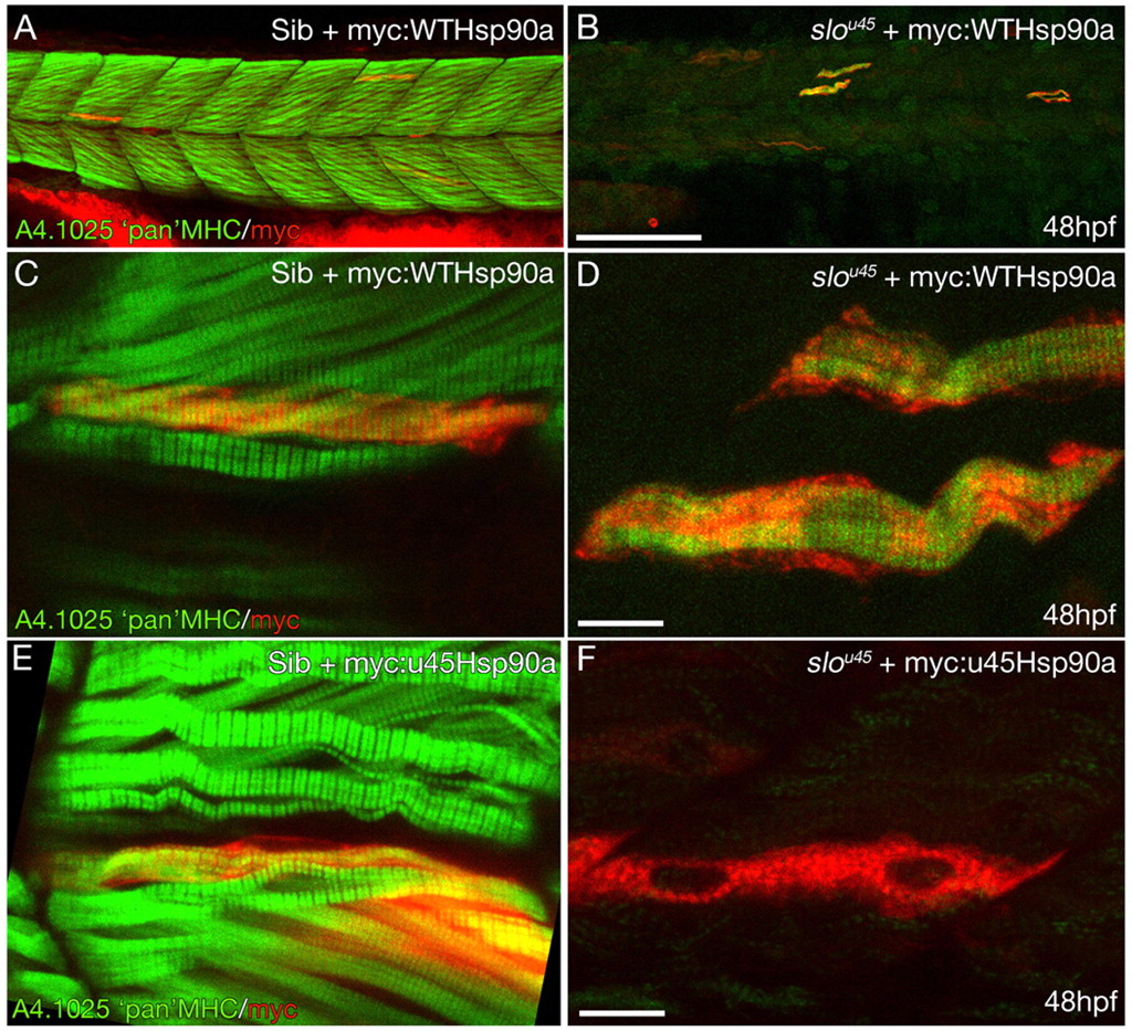
Fig. 4 Wild-type Hsp90a rescues thick filament formation in slou45 muscle cells whereas Hsp90au45 does not. (A-D) Muscle fibres from a slou45 mutant (B,D) and wild-type (Sib) (A,C) injected with DNA encoding myc:WTHsp90a double stained for myc (red) and MHC (green). MHC staining in siblings decorates the cells with organised bands (C) indicative of mature A-bands. In the slou45 mutant, striations in the MHC staining are only evident in myc+ cells (D). (E,F) Muscle fibres in sibling embryos injected with myc-tagged hsp90au45 DNA show organised MHC staining of myc+ cells (E). Myc+ cells in slou45 mutants injected with the same construct do not show organised myofibrils (F). Scale bars: 100 µm in A,B; 10 μm in C-F.
Image
Figure Caption
Acknowledgments
This image is the copyrighted work of the attributed author or publisher, and
ZFIN has permission only to display this image to its users.
Additional permissions should be obtained from the applicable author or publisher of the image.
Full text @ Development

