
IMAGE
Fig. S11
- ID
- ZDB-IMAGE-080325-119
- Publication
- Lachnit et al., 2008 - Alterations of the cytoskeleton in all three embryonic lineages contribute to the epiboly defect of Pou5f1/Oct4 deficient MZspg zebrafish embryos
- All Figures
- Figures for Lachnit et al., 2008
Image
|
Figure Caption
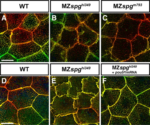
Fig. S11 Comparison and rescue of the EVL lamellipodia phenotype. Animal pole view confocal z-projections of Alexa488-Phalloidin stained EVL cells at 90% epiboly, depicted using the depth coding method. (A) WT; (B) MZspghi349; (C) MZspgm793. Extensive lamellipodia formation is observed in both alleles. (D–F) Depicts a rescue experiment showing (D) WT, (E) MZspghi349 non-injected, and (F) MZspghi349 injected with 50 pg pou5f1 mRNA. Genetically MZspghi349 mutant embryos are completely rescued by pou5f1 mRNA injection with respect to their EVL lamellipodia phenotype. Scale bar: 20 μm.
Figure Data
Acknowledgments
This image is the copyrighted work of the attributed author or publisher, and
ZFIN has permission only to display this image to its users.
Additional permissions should be obtained from the applicable author or publisher of the image.
Reprinted from Developmental Biology, 315(1), Lachnit, M., Kur, E., and Driever, W., Alterations of the cytoskeleton in all three embryonic lineages contribute to the epiboly defect of Pou5f1/Oct4 deficient MZspg zebrafish embryos, 1-17, Copyright (2008) with permission from Elsevier. Full text @ Dev. Biol.