
|
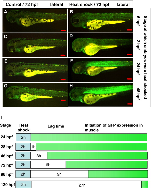
Fig. 3 Heat shock induction of GFP expression in muscle in hsp27-gfp embryos at different stages. Hsp27-gfp embryos were heat shocked for 2 h at different stages as indicated on the right and GFP expression was observed at 72 hpf. (A,C,E,G) Control untreated embryos. (B,D,F,H) Heat shock treated embryos. No GFP expression was observed in muscles of the embryos that had been heat shocked at 6 hpf (B) and 12 hpf (D). However, strong GFP expression in muscle was observed in embryos heat shocked at 24 hpf (F) and 48 hpf (H). Scale bars, 200 μm. (I) Schematic diagram showing the interval between the end of heat shock treatment and the initiation time of muscle GFP expression. Heat shock duration, lag time and the time of GFP expression in muscle are represented by blue, white and green colors.
Reprinted from Gene, 408(1-2), Wu, Y.L., Pan, X., Mudumana, S.P., Wang, H., Kee, P.W., and Gong, Z., Development of a heat shock inducible gfp transgenic zebrafish line by using the zebrafish hsp27 promoter, 85-94, Copyright (2008) with permission from Elsevier. Full text @ Gene