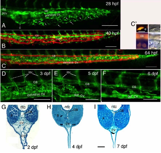
Fig. S2 Evolution of the Caudal Vein Plexus Fluorescence images of live [fli1:gfp] (A, D-F) or [fli1:gfp; gata1:dsred] (B,C) transgenic embryos, allowing visualization of endothelial cells in green, and of the flow of circulating erythrocytes - plus some progenitors in the hematopoietic mesenchyme, in red. (A) Short after the onset of blood circulation, the still single-channel caudal vein (′primitive cv′) is sprouting ventralwards all along its length. (B) By 40 hpf, blood cells flow through the multi-channel CV plexus, while by 64 hpf (C,C′) they no longer do, but the endothelial cells are still present (green). (C′) shows by comparing DIC and fluorescence images that the DsRed-positive cells present by then between CA and definitive CV are hematopoietic progenitors; two out of the three visible here appear DsRed-positive (arrows), yet to a lower extent than primitive erythrocytes, one of which is shown above for comparison (e). (D-F) Further evolution of the CV plexus-derived vascular elements, notably to connect intersomitic veins (isv) to the definitive CV. (G-I) Toluidine Blue stained cross sections of the tail, ventral to the notochord, at 2 dpf (G), 4 dpf (H) and 7 dpf (I). All bars, 100μm, except (E, G-I), 50 μm, and (C′), 10 μm.
Reprinted from Immunity, 25(6), Murayama, E., Kissa, K., Zapata, A., Mordelet, E., Briolat, V., Lin, H.F., Handin, R.I., and Herbomel, P., Tracing Hematopoietic Precursor Migration to Successive Hematopoietic Organs during Zebrafish Development, 963-975, Copyright (2006) with permission from Elsevier. Full text @ Immunity

