
|
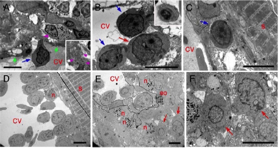
Fig. S4 Ultrastructural Organization of the Caudal Hematopoietic Tissue Supplement to Figure 3. (A,B) 5 dpf. Early lymphocyte-like (blue arrows) or myeloblast-like (red arrow) precursors in the CV lumen, one of them (A) crossing (purple arrowheads, see also inset) between CV lumen and abluminal mesenchyme through endothelium (green arrows); (B, inset) thrombocyte in the CV lumen. (C) 7 dpf, early lymphocyte-like precursor (blue arrow) in abluminal position, between endothelial cells and connective tissue. (D-F) 14 dpf; (D) Proerythroblasts, and one neutrophil (n) in the CV lumen, often adherent to its dorsal wall. (E) Granulopoiesis is still active in the mesenchyme, which contains myeloblasts (arrows), neutrophils (n), and eosinophils (eo); endothelial cell limits are delineated; (F) two myeloblasts (arrows) magnified from panel E. All bars, 5 μm
Reprinted from Immunity, 25(6), Murayama, E., Kissa, K., Zapata, A., Mordelet, E., Briolat, V., Lin, H.F., Handin, R.I., and Herbomel, P., Tracing Hematopoietic Precursor Migration to Successive Hematopoietic Organs during Zebrafish Development, 963-975, Copyright (2006) with permission from Elsevier. Full text @ Immunity