
|
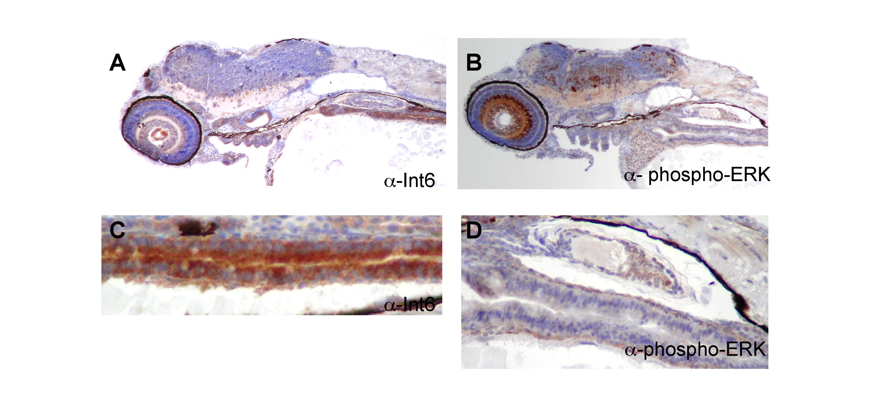
Fig. S4 Immunohistochemistry of Int6 and phospho-Erk staining in 4 dpf embryos. (A, B) While Int6 and phospho-Erk signaling overlap in the craniofacial region, they also have distinct patterns, for example in the eye and (C, D) gut. We note that while Int6 and phospho-Erk have overlapping domains of expression in the craniofacial region, Int6 staining in the craniofacial region was stronger than phospho-Erk, and phospho-Erk staining was limited to specific tissues within the craniofacial region. M, Meckel′s; E: Ethmoid plate; CH, ceratohyal; CB, ceratobrancial. Sagittal section, anterior to the left.