
|
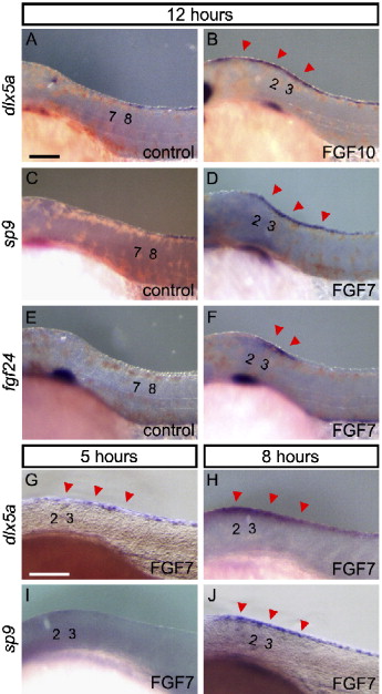
Fig. 7 Expression of marker genes for the median fin fold in ectopic structure formation induced by FGF7/10. Expression of genes in control bead-implanted embryos (A, C, E) and FGF7/10 bead-implanted embryos (B, D, F) at 12 h after implantation of beads. (A, B) dlx5a, (C, D) sp9, dlx5a (B), and sp9 (D) are expressed in the ectopic structure, continuously from the normal domain. (E, F) fgf24, which is normally expressed only in the tail bud, is (F) also seen ectopically but restricted to the epidermis around the bead. (G–J) Onset of the expression of dlx5a and sp9; dlx5a expression was detected from 5 h after implantation of beads (G, H), while sp9 expression was not detected at 5 h after (I) but was detected at 8 h after implantation of beads (J). Red arrowheads indicate an additional domain of gene expression. Numbers indicate the somite number. Scale bars in panels A and G are 100 μm.
Reprinted from Developmental Biology, 304(1), Abe, G., Ide, H., and Tamura, K., Function of FGF signaling in the developmental process of the median fin fold in zebrafish, 355-366, Copyright (2007) with permission from Elsevier. Full text @ Dev. Biol.