
Fig. 5
- ID
- ZDB-IMAGE-071109-5
- Publication
- Hall et al., 2007 - The zebrafish lysozyme C promoter drives myeloid-specific expression in transgenic fish
- All Figures
- Figures for Hall et al., 2007
|
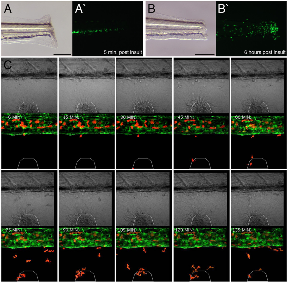
Fig. 5 Labeled cells within transgenic larvae exhibit robust responses to acute inflammation. (A and B) Inflammation assay within 7 dpf lysC::EGFP larva imaged 5 min. and 6 hours post insult, respectively. Lateral views of transected tails, anterior to left. (A/B and A'/B') Bright field and dark field images, respectively. (C) Higher resolution analysis of inflammatory response within 6 dpf lysC::DsRED2/fli1::EGFP compound transgenic animals following wounding of the ventral fin. Time-lapse images every 15 minutes (starting 6 minutes following wounding) demonstrating progressive accumulation of marked cells at the injury site. White polygon demarcates injury boundaries. Scale bars: 200 μm in A and B.