
Fig. 2
- ID
- ZDB-IMAGE-071102-8
- Genes
- Publication
- Hadzhiev et al., 2007 - Hedgehog signaling patterns the outgrowth of unpaired skeletal appendages in zebrafish
- All Figures
- Figures for Hadzhiev et al., 2007
|
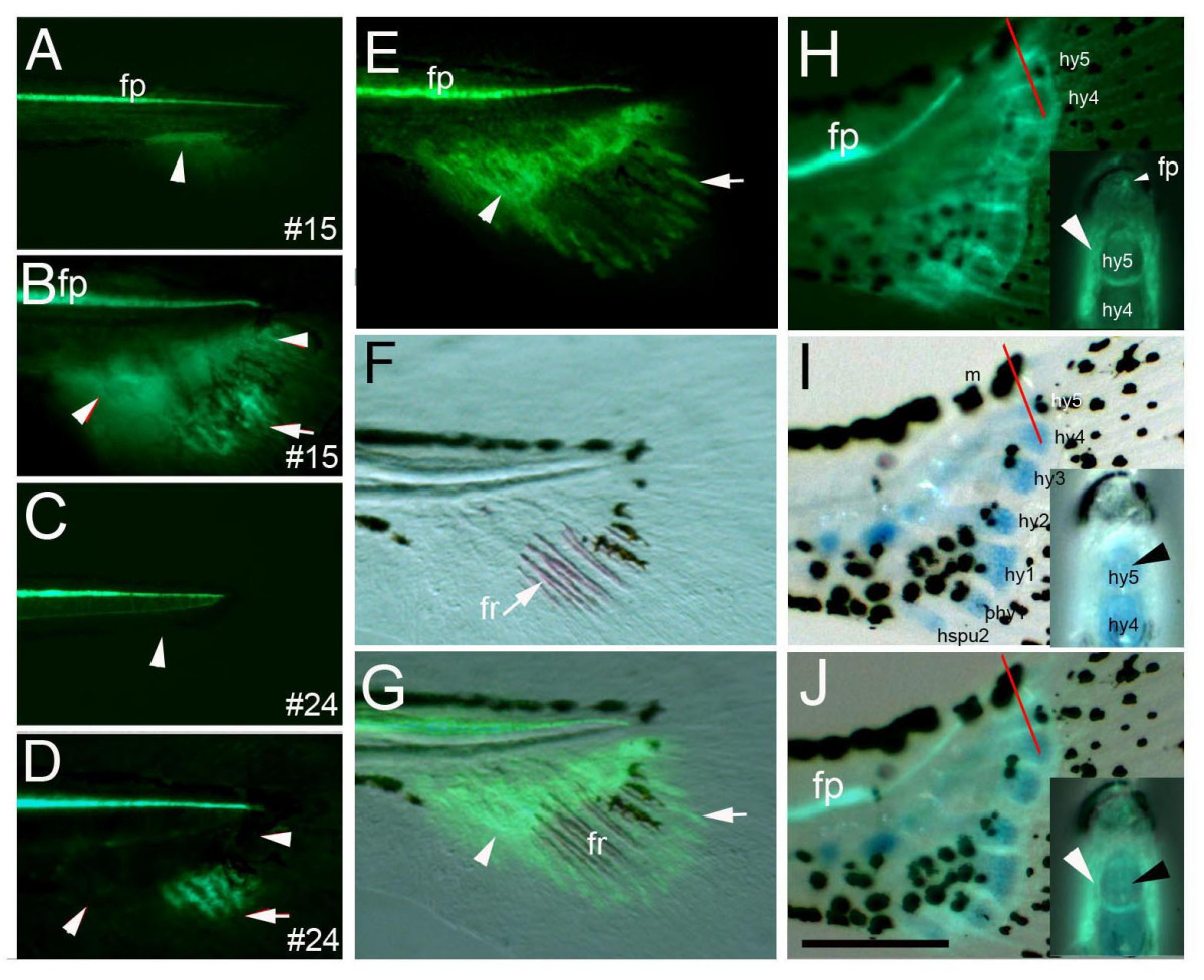
Fig. 2 The proximal GFP domain in the ACFP is independent of the fin rays and marks endoskeletal structures. Proximal GFP labeling the ACFP and its derivates (arrowheads in A, B) is only observed in one transgenic line (#15) out of 29 lines produced (compare areas with arrowheads in A to C and B to D), while GFP activity in the distal fin rays is present in several transgenic lines containing the shh regulatory elements (arrows in B, D) mimicking endogenous shh expression of the fin ray tips [21]. E-G, Proximal GFP in ACFP (arrowhead) and distal GFP domains in the fin ray tips (arrow) are physically separated during ossificiation of fin rays. Fluorescent view (E), bright field view (F) and overlay of E and F (G) of caudal fins of 12 dpf larva of transgenic line #15. F, ossification of fin rays (fr, arrow) is detected by alizarin red staining. H-J, GFP is detected in the perichondrium (arrowhead) around, but not in the endoskeletal cartilage of the hypurals marked by alcian blue staining (arrowhead). H, fluorescent view, I, bright field view, J, overlay of H and I of 14 dpf larva (5.5 mm notochord length). Red bars indicate plane of cross sections inserted in H-J. Scale bar in I indicates 120 μm (H-J). Abbreviations: fp, floor plate, n, notochord, hspu, hemal spine, phy, parhypural, hy, hypural, fr, fin ray, m, melanophore. Anatomical structures were identified as described in [19].