
Fig. S2
- ID
- ZDB-IMAGE-071102-19
- Genes
- Publication
- Hadzhiev et al., 2007 - Hedgehog signaling patterns the outgrowth of unpaired skeletal appendages in zebrafish
- All Figures
- Figures for Hadzhiev et al., 2007
|
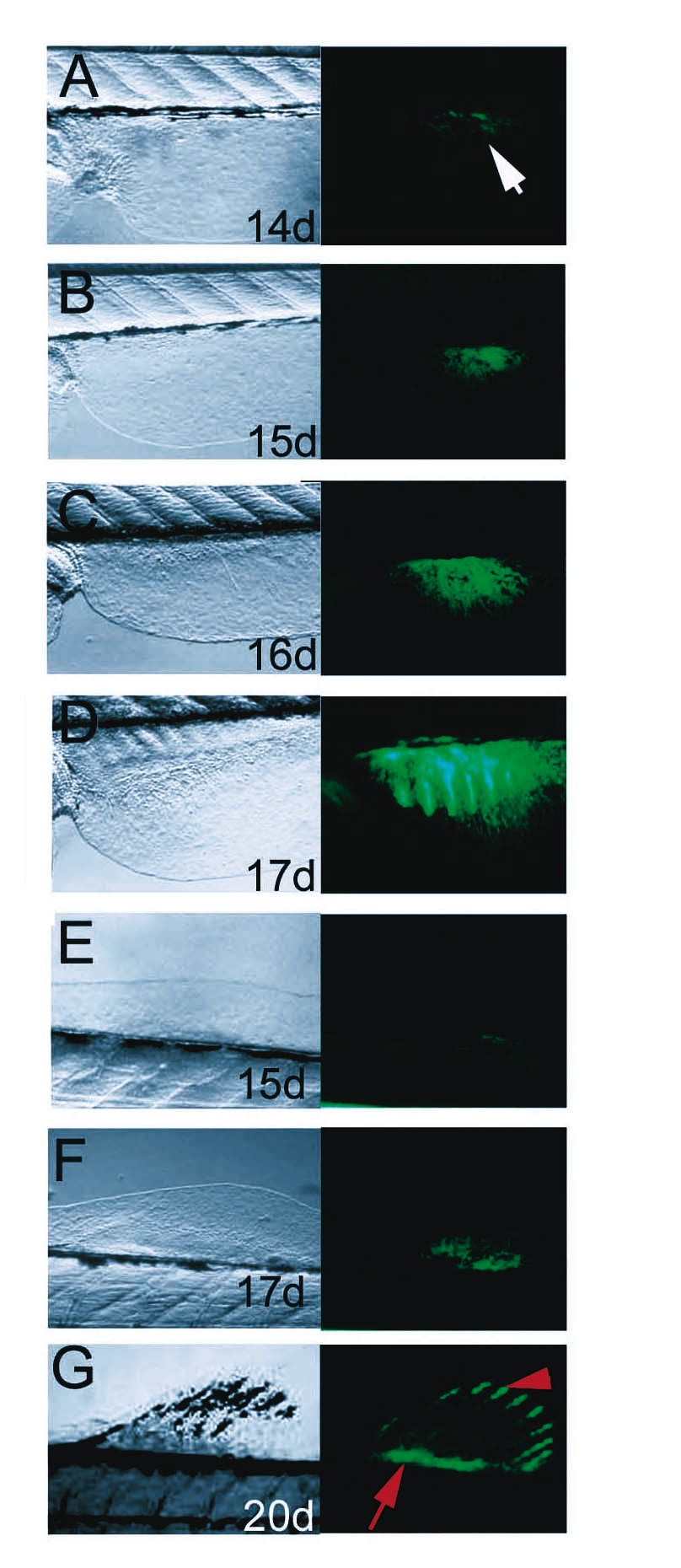
Fig. S2 Development of dorsal and anal median fins of the zebrafish. Left side panels show bright field view of dorsal fin, right panels show fluorescence signals of GFP activity. A-D: Ontogeny of the dorsal fin is marked by GFP activity. First signal is observable at 14 dpf. E, G: Development of the anal fin. GFP activity is first detected at 15 dpf. The GFP territory expands and splits into domains of the endoskeletal mesenchyme (arrow in G) and the fin ray tip (arrowhead in G) similarly to the caudal fin. H-K: Proximal expression of GFP in differentiating caudal fin primordium (arrowheads in H, I) is only observed in transgenic line #15, but not in line #24 (arrowheads in J, K), while GFP activity in the distal fin rays is present in several transgenic lines containing the shh regulatory elements (arrows in I, K) mimicking endogenous shh expression of the fin rays [21]. Age of larvae is indicated in days post fertilization (d).