
Fig. 7
- ID
- ZDB-IMAGE-071019-13
- Publication
- Yin et al., 2007 - Convergence and extension movements affect dynamic notochord-somite interactions essential for zebrafish slow muscle morphogenesis
- All Figures
- Figures for Yin et al., 2007
|
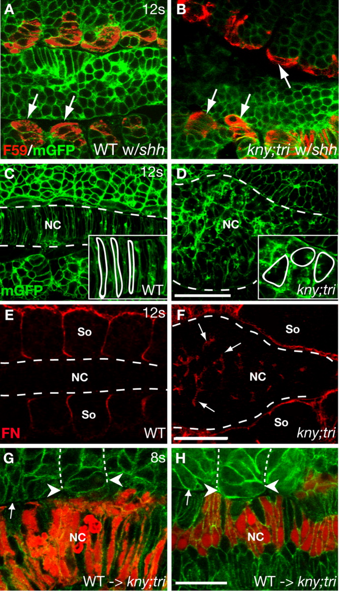
Fig. 7 Abnormal notochord properties impede the adaxial cell shape changes in kny;tri double mutants. A,B: Confocal images of the shield-depleted embryos at the 12-somite stage (15 hpf). The adaxial cells (arrows) are recognized based on F59 antibody staining. C,D: WT and kny;tri double mutant embryos expressing mGFP at the 12-somite stage. Insets show the notochord cells under higher magnification. E,F: Expression of Fn protein at the 12-somite stage (15 hpf). Dashed lines delineate the notochord-somite boundaries. Arrows in F point to the ectopic Fn expression inside the notochord in the double mutants. G,H: The presence of WT notochord cells suppressed the rotation defects of the double mutant adaxial cells lying next to them (white arrowheads). The WT donor cells are labeled with Rhodamine dextran (red). β-catenin antibody (green), which labels the cell membrane, and delineates the somite and notochord structures. A-H: Dorsal views, anterior to the left. NC, notochord; So, somite. Scale bars = 50 μm (A-F), 20 μm (G,H).