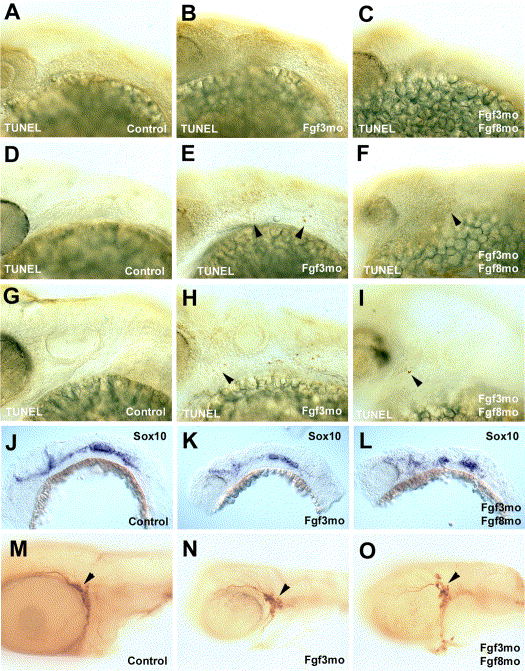
Fig. 8 Effects of inhibition of Fgf3 or Fgf3 and Fgf8 on cranial neural crest cells. (A–I) Injection of Fgf3mo either singly or in combination with Fgf8mo causes little change in cell death in pharyngula stage embryos. Dead cells are extremely rare in the arch regions of embryos injected with control morpholinos at 25 (A), 30 (D), and 48 hpf (G). Injection of Fgf3mo or both Fgf3mo and Fgf8mo results in very little cell death in the arch region at 25 hpf (B, C), while more dead cells (arrowheads) are detected at 30 (E, F) and 48 hpf (H I). (J–O) Fgf3 and Fgf8 are not essential for nonectomesenchymal neural crest cells to express Sox10 or for formation of the trigeminal ganglion. (J–L) Neural crest cells which do not form cartilage express Sox10 in 24 hpf embryos injected with either control morpholinos (J), Fgf3mo (K), or both Fgf3mo and Fgf8mo (L). (M–O) Formation of the trigeminal ganglion (arrowheads) is unimpeded in embryos injected with either control morpholinos (M), Fgf3mo (N), or both Fgf3mo and Fgf8mo (O).
Reprinted from Developmental Biology, 264(2), Walshe, J. and Mason, I., Fgf signalling is required for formation of cartilage in the head, 522-536, Copyright (2003) with permission from Elsevier. Full text @ Dev. Biol.

