
Fig. 7
- ID
- ZDB-IMAGE-071005-75
- Publication
- Matthews et al., 2004 - The zebrafish onecut gene hnf-6 functions in an evolutionarily conserved genetic pathway that regulates vertebrate biliary development
- All Figures
- Figures for Matthews et al., 2004
|
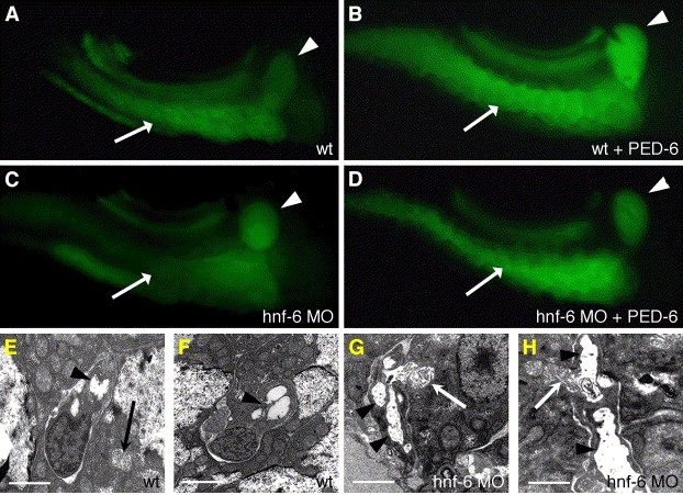
Fig. 7 Reduced biliary lipid secretion and cholestasis in hnf-6 morphants. (A–D) Fluorescent images of 6 dpf larvae (right lateral view). (A and C) Endogenous, baseline gallbladder (arrowhead) and intestinal (arrow) fluorescence in 6 dpf WT (A) and hnf-6 morphant (C) larvae that did not receive the PED-6 lipid reporter. (B and D) Identical views of 6 dpf sibling larvae soaked overnight in PED-6. Note enhanced gallbladder fluorescence in WT (B) but not in the hnf-6 morphant (D). These images are representative of 20 paired sets of larvae examined. (E–H) Liver ultrastructure in 5 dpf WT (E and F) and hnf-6 morphant (G and H) larvae. Arrowheads point to lumen of the capillary-like ductules in the WT specimen. Arrows in all of these electron micrographs point to adjacent hepatocyte canaliculi. Ductular lumens in hnf-6 morphants (arrowheads) are enlarged and ectatic compared with WT. Scale bars = 2 μm.
Reprinted from Developmental Biology, 274(2), Matthews, R.P., Lorent, K., Russo, P., and Pack, M., The zebrafish onecut gene hnf-6 functions in an evolutionarily conserved genetic pathway that regulates vertebrate biliary development, 245-259, Copyright (2004) with permission from Elsevier. Full text @ Dev. Biol.