
Fig. 7
- ID
- ZDB-IMAGE-071004-94
- Genes
- Publication
- Hernandez-Lagunas et al., 2005 - Zebrafish narrowminded disrupts the transcription factor prdm1 and is required for neural crest and sensory neuron specification
- All Figures
- Figures for Hernandez-Lagunas et al., 2005
|
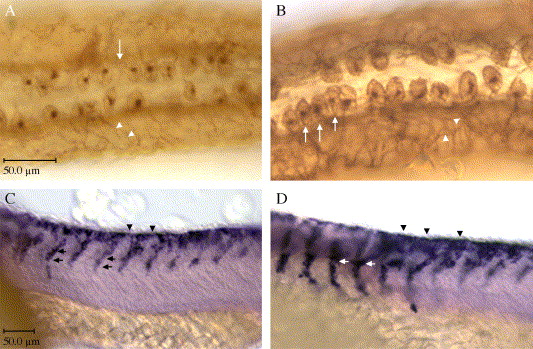
Fig. 7 Ectopic expression of prdm1 increases neural crest and Rohon–Beard sensory neurons. (A, B) HNK-1 immunohistochemistry at 24 hpf; dorsal views, anterior to the left. (A) Wildtype expression of HNK-1, (B) Subsequent to prdm1 mRNA injections, RB sensory neurons are increased (arrows). (C, D) Crestin expression at 24 hpf, lateral views, anterior to the left. (C) Wildtype expression of crestin, (D) Following injection of prdm1 mRNA, the neural crest emigration and migration streams appear thick with cells (arrows) as compared with the same region in wildtype embryos. The migration of the cells also appears premature in the prdm1 expressing embryos. At the same time point in development, cells in prdm1-expressing embryos have migrated down to the yolk, whereas wildtype cells are only half way down their migration path. Scale bar for A, B is in A and is 50 μm; for C, D the scale bar is in C and is 50 μm.
Reprinted from Developmental Biology, 278(2), Hernandez-Lagunas, L., Choi, I.F., Kaji, T., Simpson, P., Hershey, C., Zhou, Y., Zon, L., Mercola, M., and Artinger, K.B., Zebrafish narrowminded disrupts the transcription factor prdm1 and is required for neural crest and sensory neuron specification, 347-357, Copyright (2005) with permission from Elsevier. Full text @ Dev. Biol.