
|
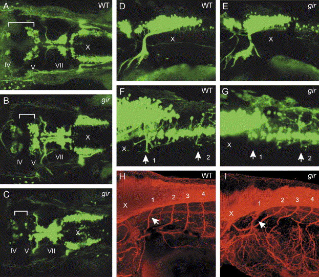
Fig. 8 Patterning of motor neurons is affected in the gir mutant. (A–G) Expression of the Isl1-GFP transgene was examined by fluorescence microscopy. (A–E) Dorsal views of hindbrain (A–C) and lateral views of caudal hindbrain and rostral spinal cord (D and E) in wild type (A and D) and gir mutant (B, C, and E) embryos at 48 hpf. The distance between the trochlear (nIV) and trigeminal (nV) nuclei is reduced, and bilateral clusters of the vagal nucleus (nX) are broadened, in the gir mutant (B and C). In some of the gir mutants, the number of neurons in the trigeminal nucleus (nV) is reduced (C). (F and G) Lateral views of caudal hindbrain and rostral spinal cord in wild type (F) and gir mutant (G) embryos at 3 dpf. Dorsal projection of motor neuron axons in the rostralmost spinal cord (labeled by arrow 1) is absent in the gir mutant (G). (H and I) Immunostaining of motor neurons with acetylated α-tubulin antibody in the caudal hindbrain and rostral spinal cord of wild type (H) and gir mutant (I) embryos at 3 dpf. Axons of spinal motor neurons are not significantly affected in the gir mutant (I). Arrows indicate axons from the rostralmost spinal motor neurons.
Reprinted from Developmental Biology, 278(2), Emoto, Y., Wada, H., Okamoto, H., Kudo, A., and Imai, Y., Retinoic acid-metabolizing enzyme Cyp26a1 is essential for determining territories of hindbrain and spinal cord in zebrafish, 415-427, Copyright (2005) with permission from Elsevier. Full text @ Dev. Biol.