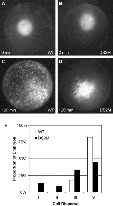
Fig. 8 Dscam is required for cell dispersal during epiboly. (A) Wild-type embryo at high cell stage showing bright spot of FITC-dextran labeled cells after uncaging. (B) DS2M injected embryo at high cell stage showing a similar size spot of FITC-dextran labeled cells. (C) A wild-type embryo imaged after 2 h of development illustrating how cells within the original spot widely disperse across the embryo. This embryo is an example of Grade IV dispersal. (D) A DS2M embryo imaged 2 h after uncaging shows most cells contained within the original spot. Limited migration from the spot has occurred as the border of the original spot is irregular. This is an example of Grade I dispersal. (E) Microinjection of DS2M (n = 36) shifts population of embryos exhibiting less dispersal of cells within the uncaged spot toward Grade I as compared to wild-type embryos (n = 28). This shift is statistically significant by χ2, P = 0.01. Grade I, uncaged spot is condensed with slight cell migration. Grade II, spot is slightly dispersed. Grade III, spot is moderately dispersed. Grade IV, spot is widely dispersed.
Reprinted from Developmental Biology, 279(1), Yimlamai, D., Konnikova, L., Moss, L.G., and Jay, D.G., The zebrafish down syndrome cell adhesion molecule is involved in cell movement during embryogenesis, 44-57, Copyright (2005) with permission from Elsevier. Full text @ Dev. Biol.

