
Image
|
Figure Caption
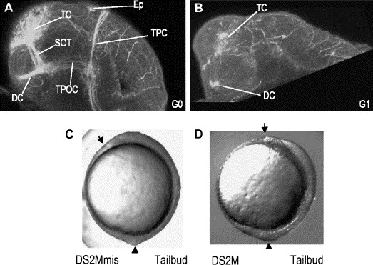
Fig. 5 dscam morphants show axon tract defects. (A) G0 embryo shows normal axon tracts. (B) G1 embryo shows few neurons and disorganized axons. Ep, epiphysis; DC, diencephalon; SOT, supraoptic tract; TC, telencephalon; TPOC, tract of the post-optic commissure; TPC, tract of the posterior commissure. (C) Control and (D) dscam knockdown embryos at tailbud stage. Arrowhead represents tailbud. Arrow represents leading edge of the polster.
Acknowledgments
This image is the copyrighted work of the attributed author or publisher, and
ZFIN has permission only to display this image to its users.
Additional permissions should be obtained from the applicable author or publisher of the image.
Reprinted from Developmental Biology, 279(1), Yimlamai, D., Konnikova, L., Moss, L.G., and Jay, D.G., The zebrafish down syndrome cell adhesion molecule is involved in cell movement during embryogenesis, 44-57, Copyright (2005) with permission from Elsevier. Full text @ Dev. Biol.