
|
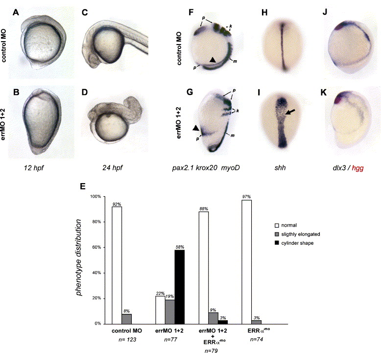
Fig. 4 Effect of ERRα-targeting MOs on zebrafish later development. (A–D). Lateral views of living embryos (control MO- (A,C) or errMO- (B,D) injected) at 12 hpf (A–B, dorsal to the right) and at 24 hpf (C–D). (E) Strength of the observed phenotypes. Injected embryos were scored at 12 hpf as unaffected, caudally elongated or cylinder shaped. Number of embryos examined for each condition is indicated below the graphe. (F–K) In situ hybridization with a mix of myoD (m), pax 2.1 (p) and krox20 (k) probes (F–G, lateral views, dorsal to the right), shh probe (H–I, dorsal views) or hgg (in red) and dlx3 (J–K, lateral views, dorsal to the right), on embryos at 12 hpf, injected either with control MO (F, H, J) or errα targeting MO (G, I, K). Note the pronephric ducts (F–G, arrowheads) in a ventral position in errMO-injected animals. shh expression domain (I, arrow) is enlarged, but less dense, in the morphant as compared to control. All embryos presented here did not display a strong delay in epiboly.
Reprinted from Developmental Biology, 281(1), Bardet, P.L., Horard, B., Laudet, V., and Vanacker, J.M., The ERRalpha orphan nuclear receptor controls morphogenetic movements during zebrafish gastrulation, 102-111, Copyright (2005) with permission from Elsevier. Full text @ Dev. Biol.