
Fig. 2
- ID
- ZDB-IMAGE-071001-99
- Genes
- Publication
- Te Velthuis et al., 2007 - Gene expression patterns of the ALP family during zebrafish development
- All Figures
- Figures for Te Velthuis et al., 2007
|
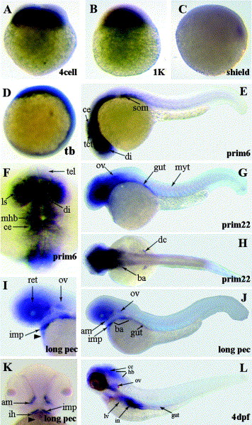
Fig. 2 Expression pattern of the zebrafish alp gene by whole mount in situ hybridization. Zebrafish embryos at eight different developmental stages are shown and stages were indicated. Expression domains are marked by arrows and expression was found in the following structures: adductor mandibulae (am), branchial arches (ba), cerebellum (ce), diencephalon (di), duct of Cuvier (dc), hindbrain (hb), interhyoideus (ih), intermandibularis posterior (imp), intestine (in), lens (ls), liver (lv), midbrain-hindbrain boundary (mhb), myotomes (myt), otic vesicle (ov), retina (ret), somites (som), tectum (tct), telencephalon (tel). Black arrowheads point to the developing heart.
Reprinted from Gene expression patterns : GEP, 7(3), Te Velthuis, A.J., Ott, E.B., Marques, I.J., and Bagowski, C.P., Gene expression patterns of the ALP family during zebrafish development, 297-305, Copyright (2007) with permission from Elsevier. Full text @ Gene Expr. Patterns