
Image
|
Figure Caption
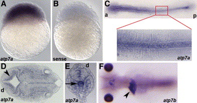
Fig. 5 Expression pattern of atp7a via in situ hybridization
Anterior (a), posterior (p), and dorsal (d) are as indicated.
A) atp7a message is present in all cells at 3 hpf.
B) Staining is specific, as a sense-control probe does not show staining.
C) At 8 somites, atp7a is expressed most abundantly in the notochord and is expressed ubiquitously at lower levels.
D and E) Sectioning at 24 hpf reveals staining along the ventricle (D) (arrowhead) and in the distal notochord (E) (arrow).
F) In situ analysis of atp7b reveals expression in the liver (arrowhead) in a 5 dpf embryo as shown in this dorsal view.
Figure Data
Acknowledgments
This image is the copyrighted work of the attributed author or publisher, and
ZFIN has permission only to display this image to its users.
Additional permissions should be obtained from the applicable author or publisher of the image.
Reprinted from Cell Metabolism, 4(2), Mendelsohn, B.A., Yin, C., Johnson, S.L., Wilm, T.P., Solnica-Krezel, L., and Gitlin, J.D., Atp7a determines a hierarchy of copper metabolism essential for notochord development, 155-162, Copyright (2006) with permission from Elsevier. Full text @ Cell Metab.