
IMAGE
Fig. 6
- ID
- ZDB-IMAGE-071001-103
- Genes
- Publication
- Te Velthuis et al., 2007 - Gene expression patterns of the ALP family during zebrafish development
- All Figures
- Figures for Te Velthuis et al., 2007
Image
|
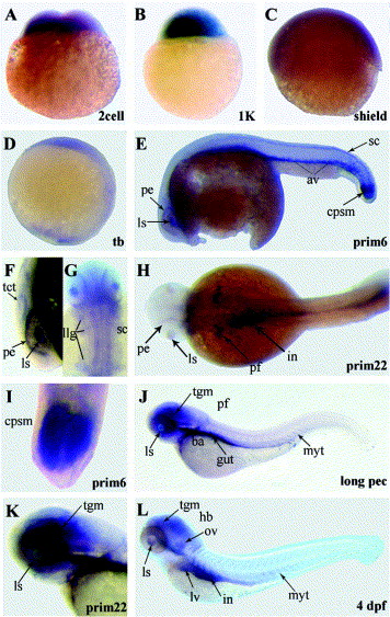
Figure Caption
Fig. 6 Expression pattern of the zebrafish ril gene by in situ hybridization during zebrafish embryonic development. ril expression was found in the branchial arches (ba), caudal presomitic mesoderm (cpsm), hindbrain (hb), intestine (in), lateral line ganglia (llg), lens (ls), liver (lv), myotomes (myt), pigmented epithelium (pe), pectoral fin (pf), spinal cord (sc), somites (som), tectum (tct), telencephalon (tel) and finally the tegmentum (tgm).
Figure Data
Acknowledgments
This image is the copyrighted work of the attributed author or publisher, and
ZFIN has permission only to display this image to its users.
Additional permissions should be obtained from the applicable author or publisher of the image.
Reprinted from Gene expression patterns : GEP, 7(3), Te Velthuis, A.J., Ott, E.B., Marques, I.J., and Bagowski, C.P., Gene expression patterns of the ALP family during zebrafish development, 297-305, Copyright (2007) with permission from Elsevier. Full text @ Gene Expr. Patterns