
Fig. 2
- ID
- ZDB-IMAGE-070928-1
- Publication
- Hirata et al., 2007 - Zebrafish relatively relaxed mutants have a ryanodine receptor defect, show slow swimming and provide a model of multi-minicore disease
- All Figures
- Figures for Hirata et al., 2007
|
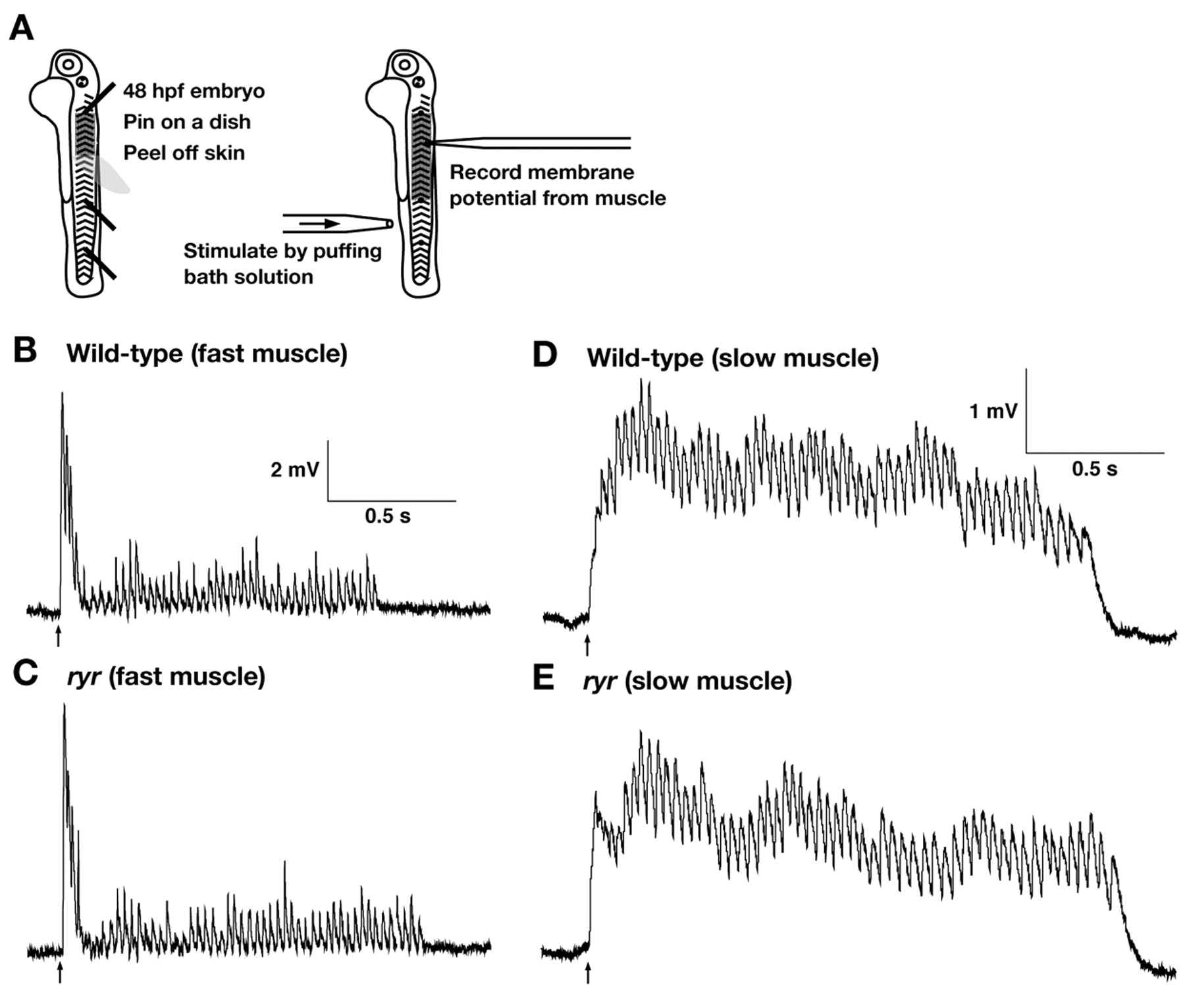
Fig. 2 The CNS and NMJ function normally in ryr mutants. (A) Schematic summary of the experimental procedure. Embryos (48 hpf) are pinned on a dish with tungsten wires through the notochord so as not to damage the CNS. The skin is peeled off to allow access to the muscle cells. Muscle voltage responses are evoked by mechanosensory stimulation delivered by a puff of bath solution to the tail and are recorded with a patch electrode. (B,C) Voltage recordings from fast muscle fibers of a wild-type sibling (B) and an ryr mutant (C) displayed similar patterns of depolarizations (fictive swimming). (D,E) Voltage recordings from slow muscle of a wild-type sibling (D) and an ryr mutant (E) display similar rhythmic depolarizations. Arrows indicate the time of stimulation.