
Image
|
Figure Caption
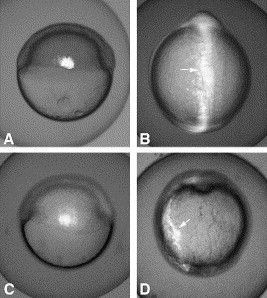
Fig. 6 Analysis of convergence in wt (A, B) and blf morphants (C, D) by uncaging DMNB-caged fluorescein-dextran in a group of cells within lateral mesendoderm. (A, C) A group of cells within lateral mesendoderm is labeled by the UV-photoactivation at the shield stage. Lateral view, dorsal is to the right. (B, D) The same embryo as in panels (A) or (C) at the 2-somite stage. Note that the labeled cells (arrows) have converged to the midline in the control (B) but not in the morphant embryo (D). Dorsal view, anterior is up.
Acknowledgments
This image is the copyrighted work of the attributed author or publisher, and
ZFIN has permission only to display this image to its users.
Additional permissions should be obtained from the applicable author or publisher of the image.
Reprinted from Developmental Biology, 283(1), Sumanas, S., Zhang, B., Dai, R., and Lin, S., 15-Zinc finger protein Bloody Fingers is required for zebrafish morphogenetic movements during neurulation, 85-96, Copyright (2005) with permission from Elsevier. Full text @ Dev. Biol.