
Fig. 4
- ID
- ZDB-IMAGE-070925-87
- Publication
- Albertson et al., 2007 - Fgf8 haploinsufficiency results in distinct craniofacial defects in adult zebrafish
- All Figures
- Figures for Albertson et al., 2007
|
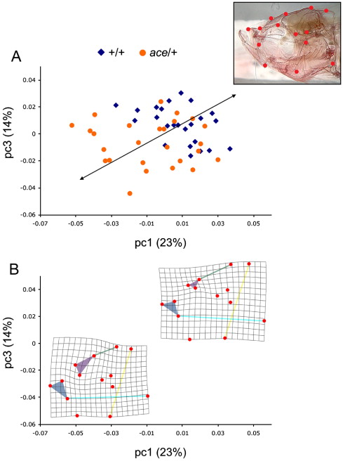
Fig. 4 Fig. 4. Aceti282a/+ zebrafish exhibit aberrant craniofacial geometry. A relative warp analysis revealed two principal component axes that discriminate aceti282a/+ zebrafish from their wild-type siblings (A). The inset illustrates the position of fourteen landmarks used to describe the craniofacial skeleton in the lateral view. (B), Deformation grids as a function of the axes that differentiate aceti282a/+ and +/+ individuals. Aceti282a/+ animals exhibited an expanded frontal region of the skull (purple triangle), relative to the length of the posterior region of the cranium (green line). Aceti282a/+ zebrafish also exhibited coordinated changes in the configuration of the upper jaws (blue triangle), and in the relative height of the skull (yellow line).
Reprinted from Developmental Biology, 306(2), Albertson, R.C., and Yelick, P.C., Fgf8 haploinsufficiency results in distinct craniofacial defects in adult zebrafish, 505-515, Copyright (2007) with permission from Elsevier. Full text @ Dev. Biol.