
Fig. 2
- ID
- ZDB-IMAGE-070925-54
- Publication
- Liu et al., 2007 - Cloning and expression analysis of cadherin7 in the central nervous system of the embryonic zebrafish
- All Figures
- Figures for Liu et al., 2007
|
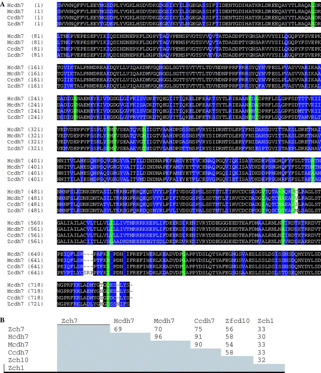
Fig. 2 (A) Amino acid sequence comparison between the deduced zebrafish Cdh7 amino acid sequence (Zcdh7), chicken Cdh7 (Ccdh7), human Cdh7 (Hcdh7), and mouse Cdh7 (Mcdh7). Comparisons were between published sequences from the EC1 to near the end of the coding sequences. Residues that are identical among all four sequences are outlined in black. Residues conserved in three of four sequences are outlined in blue, while two of four outlined in green. (B) Sequence identity percentages for pairwise comparisons between zebrafish Cdh7, several other Cdh7 sequences, cadherin10 (Zcdh10, another type II zebrafish cadherin) and a type I zebrafish cadherin, cadherin1 (Zcdh1). Sequence comparisons were performed using Clustal W (Des Higgins, EBI, Hinxton Hall, UK).
Reprinted from Gene expression patterns : GEP, 7(1-2), Liu, B., Joel Duff, R., Londraville, R.L., Marrs, J.A., and Liu, Q., Cloning and expression analysis of cadherin7 in the central nervous system of the embryonic zebrafish, 15-22, Copyright (2007) with permission from Elsevier. Full text @ Gene Expr. Patterns