
|
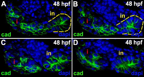
Fig. S1 Exocrine anlage architecture. (A–D) Serial 3 μm sections through the pancreas of a 48 hpf zebrafish larva processed for cadherin (green) immunohistochemistry and dapi (blue). Exocrine cells surround the islet, and they are adjacent to the intestine (yellow dashed line). These cadherin immunostainings show that the exocrine cells are not arranged as a simple columnar epithelium. Compare appearance of the intestinal epithelium, which in these sections is cut tangentially, to the exocrine cells. Most intestinal epithelial cells have a columnar appearance, whereas the exocrine cells appear round or have a stellate appearance and they are stratified. cad: cadherin; i: islet; in: intestine.
Reprinted from Developmental Biology, 284(1), Yee, N.S., Lorent, K., and Pack, M., Exocrine pancreas development in zebrafish, 84-101, Copyright (2005) with permission from Elsevier. Full text @ Dev. Biol.